



Antibody recognition depends on more than the amino acid sequence of a binding region. It emerges from the three-dimensional arrangement of residues that form an interface with an antigen. Antibody CDR design aims to modify or generate complementarity-determining region (CDR) sequences that support a desired interaction, while preserving the structural context needed for binding.
As artificial intelligence expands the possibilities for antibody engineering, understanding how CDRs, framework regions, and heavy–light chain pairing shape antigen recognition remains essential for developing candidates that perform experimentally.
Conventional antibodies contain two heavy chains and two light chains. Each antigen-binding site is formed by a heavy-chain variable domain (VH) paired with a light-chain variable domain (VL).
Together, these domains contain six CDRs: HCDR1, HCDR2, and HCDR3 from the heavy chain, and LCDR1, LCDR2, and LCDR3 from the light chain. These loops are supported by relatively conserved framework regions that help maintain their structural organization.
The variable domains form the recognition surface, while the complete antibody also contains constant domains involved in stability and effector functions. Single-domain antibodies, including VHHs, differ by using one variable domain containing three CDRs.
The paratope is the antibody surface that interacts with an antigen, while the epitope is the corresponding region recognized on the antigen.
Although CDRs generally provide much of the binding interface, their annotated boundaries do not precisely define the paratope. Not every CDR residue contacts the antigen, and some framework residues can participate directly in recognition.
Antigen recognition depends on the shape and chemical properties of the interacting surfaces. Hydrogen bonds, electrostatic interactions, hydrophobic contacts, and van der Waals interactions collectively influence binding.
Residues from different CDRs may form a shared interface, although their contributions vary between antibodies. HCDR3 frequently plays a prominent role because of its substantial sequence and structural diversity, but it is not the sole determinant of specificity.
Framework residues and VH-VL orientation also influence loop positioning. Consequently, changing one CDR can affect neighboring interactions, making it important to evaluate designed sequences within their intended antibody framework and chain-pairing context.
Antibody numbering systems, including Kabat, Chothia, and IMGT, use different criteria to identify CDR boundaries. As a result, the same antibody sequence may have different residues classified as CDRs depending on the scheme.
These distinctions matter in computational design because boundary selection determines which residues a model can modify and how predictions are compared.
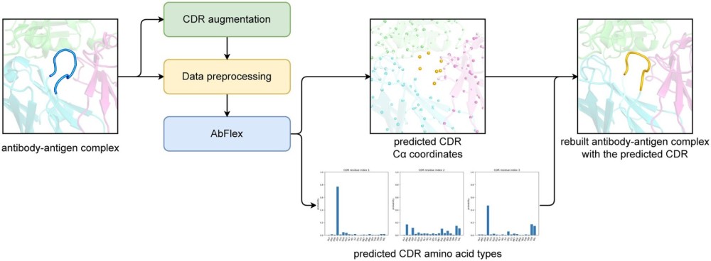
A 2024 study by Jeon and Kim, published in Bioinformatics, demonstrated how flexible CDR definitions could improve computational antibody design.1 Their approach augmented training data by shifting the positions used to define CDR segments, allowing the model to learn from different local sequence and structural contexts.
Importantly, changing computational CDR boundaries during training does not itself alter the physical antibody. Actual loop-length engineering requires sequence insertions or deletions and may introduce additional structural constraints.
Among the six CDRs, HCDR3 typically displays the greatest sequence and conformational diversity. Its variability presents both an opportunity and a challenge for AI design: it can contribute strongly to antigen recognition but is often difficult to predict accurately.
Other CDRs remain important, however. Their interactions can help stabilize the interface, influence specificity, and shape the orientation of HCDR3.
Rather than optimizing HCDR3 in isolation, antibody engineering increasingly benefits from considering the complete binding surface, including contributions from the light chain and surrounding framework.
AI-guided approaches can explore CDR sequence variants, predict compatible conformations, and prioritize candidates for experimental evaluation.
Sequence-based methods learn patterns from antibody sequences, while structure-conditioned approaches incorporate three-dimensional information about the antibody, antigen, or their complex. Other generative methods jointly propose sequences and structures.
The appropriate approach depends on the design objective, whether refining an existing antibody or developing recognition of a new epitope.
Predictions must nevertheless be interpreted cautiously. Structural plausibility and favorable computational interaction scores do not establish experimentally measured affinity or specificity.
| Design Feature | AI-Guided Evaluation | Experimental Follow-up |
|---|---|---|
| CDR sequence | Promising substitutions | Expression and binding |
| Loop geometry | Structural compatibility | Structural analysis |
| Paratope chemistry | Potential antigen contacts | Binding and specificity assays |
| Framework context | Stability and compatibility | Expression and developability |
| Overall interface | Predicted recognition | Functional testing |
Experimental validation determines whether computationally promising antibodies retain the properties required for further development.
Initial characterization should establish expression and sample quality, including concentration, purity, and aggregation where appropriate. Inconsistent sample quality can complicate comparisons between variants.
Binding measurements using ELISA, biolayer interferometry (BLI), or surface plasmon resonance (SPR) can assess recognition and, depending on the assay, binding kinetics and affinity. Specificity counterscreens and cell-based functional assays provide additional evidence where relevant.
Targeted mutagenesis can help identify residues important for recognition, although changes in binding may reflect altered folding rather than direct loss of an antigen contact.
Importantly, stronger binding does not necessarily translate into improved biological activity or developability.
CDR modifications can affect more than affinity. Changes in exposed hydrophobic residues, charge distribution, or local flexibility may influence aggregation, self-association, nonspecific binding, and stability.
Selecting candidates therefore requires balancing measured binding performance with developability characteristics rather than relying on a single computational score.
As AI models generate increasingly large panels of antibody variants, experimental characterization becomes essential for identifying candidates with useful properties.
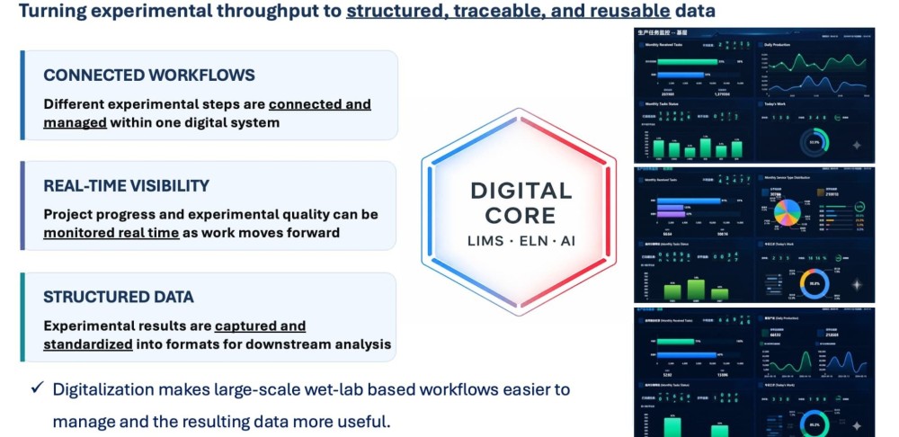
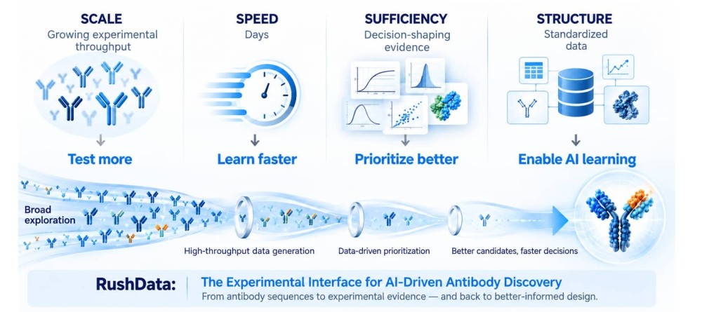
Biointron supports this process through recombinant antibody expression, affinity maturation, and developability assessment. Its RushDATA™ platform enables high-throughput characterization of designed antibody variants, generating structured experimental datasets that can support candidate comparison and iterative AI-driven discovery.
Integrating computational design with reproducible wet-lab measurements helps researchers distinguish promising predictions from experimentally validated antibodies.
Contact Biointron to discuss expression and characterization of your CDR-designed antibody variants.
The computational or experimental modification of antibody CDR sequences to influence antigen recognition.
CDRs are defined sequence regions; the paratope comprises the residues and surface involved in antigen binding.
No. Their contributions vary, and some may make little or no direct contact.
They establish residue boundaries used in sequence analysis, structural comparisons, and computational design.
No. Computational predictions require experimental validation to establish binding improvements.
Woosung Jeon, Dongsup Kim, AbFlex: designing antibody complementarity determining regions with flexible CDR definition, Bioinformatics, Volume 40, Issue 3, March 2024, btae122, https://doi.org/10.1093/bioinformatics/btae122
Learn how generative AI antibody design creates new sequences using target const……
Artificial intelligence (AI) is transforming antibody discovery by enabling rese……
AI-driven antibody discovery has made it possible to generate thousands of candi……