



Antibody discovery often begins with a search through biological diversity. Following immunization or infection, B cells generate large repertoires of antibodies with different sequences, affinities and functional properties. The challenge is finding the relatively small number of antibodies worth pursuing while preserving enough information to identify and reproduce them.
Single-cell approaches have made this process more direct by allowing antibody sequences to be recovered from individual B cells. Plasma cells are particularly interesting targets. These terminally differentiated B cells are specialized for producing and secreting antibodies and can provide access to highly matured immune repertoires. Antibodies isolated from antibody-secreting cells (ASCs), which include plasma cells and plasmablasts, are thought to have higher affinity on average than antibodies obtained from memory B cells.
Their biology, however, creates a screening problem. Plasma cells release their antibodies into the surrounding environment rather than displaying large amounts of immunoglobulin on the cell surface. As a result, conventional antigen-specific cell sorting cannot interrogate them as straightforwardly as memory B cells.
Droplet microfluidics offers one way around this limitation: isolate a cell together with the antibody it secretes, measure that antibody within a microscopic compartment, and retain the connection to the cell that carries its sequence.
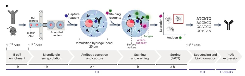
Fluorescence-activated cell sorting, or FACS, separates cells according to fluorescent signals associated with selected cellular characteristics. For antibody discovery from memory B cells, researchers can label an antigen and identify B cells whose membrane-bound immunoglobulin recognizes that antigen.
Plasma cells present a different situation. Because they secrete antibodies and express few or no immunoglobulins on their surface, their antigen specificity cannot usually be read directly from the outside of the cell. Traditional approaches such as ELISpot can identify populations secreting antigen-specific antibodies, but they do not inherently recover the antibody genotype from each positive cell.
This creates a problem in plasma-cell antibody discovery: the antibody phenotype exists outside the cell, while the antibody genes remain inside it.
Here, phenotype refers to a measurable characteristic of the secreted antibody, such as antigen binding or biological activity. Genotype refers to the genetic sequence encoding that antibody.
An effective single-cell screening method therefore needs to measure the antibody while maintaining its association with the cell that produced it.
Microfluidics is the manipulation of very small volumes of fluid through microscale channels and structures. Several microfluidic formats have been applied to antibody screening, including microvalves, micro- and nanowells, and droplets. They solve different experimental problems.
Microvalve systems can provide precise control of reagent movement but may require relatively complex device architectures. Microwells physically separate individual cells and can retain secreted molecules for analysis, although the number of cells that can be screened is constrained by the number of available wells.
Droplet-based systems take a different approach. Cells and assay reagents are encapsulated within discrete water-in-oil droplets that function as independent reaction chambers. Compared with fixed microchamber systems, droplet platforms can support much larger numbers of individual assays. Importantly for antibody discovery, molecules secreted by a cell remain confined within its droplet, maintaining a physical connection between the cell's genotype and the phenotype of its secreted product.
That confinement is particularly useful for plasma cells.
A single plasma cell may continuously release antibody molecules, but in a conventional bulk culture those molecules diffuse into a much larger volume. Determining which cell produced which antibody then becomes difficult.
A microfluidic droplet confines the secreted molecules within a volume that can be on the picoliter scale. A picoliter is one trillionth of a liter.
This reduced volume limits dilution of the secreted antibody, which can improve signal detection. A recent review of droplet microfluidics notes that picoliter compartmentalization can increase sensitivity for low-abundance hits by concentrating secreted molecules and improving signal-to-noise.1
Droplets can also be generated rapidly. Typical droplet-generation systems operate at frequencies in the kilohertz range, although actual cell-screening throughput depends on the platform, assay design, and sorting system.
Once an antigen-specific cell has been identified, single-cell sequencing can recover the antibody genes associated with that cell.
This is important because a conventional antibody contains both a heavy chain and a light chain. Native heavy-light chain pairing refers to retaining the naturally occurring heavy and light chains that were produced together by the same B cell.
Single-cell workflows can preserve this pairing because both sequences are recovered from an individual cell. By contrast, combinatorial library approaches such as phage display may disrupt the original pairing of heavy and light chains, depending on how the library is constructed.
Droplet screening can be designed around several types of experimental readouts. The most straightforward is antigen binding, but published systems have also explored assays that measure functional properties.2
In one common format, an antibody-secreting cell is encapsulated together with antigen-coated beads and fluorescent detection reagents. As the cell secretes antibody, antigen-specific molecules are captured and generate a localized fluorescent signal. Droplets above a defined fluorescence threshold can then be sorted for downstream analysis.
Binding does not necessarily reveal whether an antibody changes the biological activity of its target. Droplet assays can instead be configured around a functional readout. One early example described in the 2025 review screened hybridoma cells for antibodies that inhibited angiotensin-converting enzyme 1. The desired antibody-producing cells were initially present at a ratio of 1:10,000 within an unrelated population, and droplets were selected according to enzyme inhibition rather than binding alone.1
Microfluidic systems have also been explored for identifying antibodies that are internalized after binding to a cell-surface target. Antibody internalization occurs when an antibody binds its target at the cell surface and the resulting complex is taken into the cell. This property is especially relevant for therapeutic formats such as antibody-drug conjugates, where intracellular delivery of a payload can be important.
Other experimental systems have used droplets to distinguish viral infection from antibody-mediated neutralization. These assays incorporate viral particles, host cells or detection systems so that neutralizing activity can be associated with a measurable fluorescence signal.
Recent research illustrates that the same biological problem can be solved through different microfluidic designs.
In a recent study, Fischer and colleagues encapsulated individual antibody-secreting cells in droplets containing agarose.3 After the agarose solidified, antibody-capture reagents immobilized antibodies around the cell that secreted them. The resulting hydrogel particles could be stained, washed and sorted using conventional FACS, while retaining the connection between secreted antibody and cell of origin.
The researchers screened millions of mouse and human immune cells and recovered SARS-CoV-2 monoclonal antibodies. Among a representative subset of human antibodies, 95% bound the corresponding antigen, with many showing subnanomolar affinity and neutralizing activity below 100 ng ml−1. The workflow generated pathogen-specific antibodies within approximately two weeks.
AbDrop uses a different implementation of the same general principle: detect antigen-specific antibody secretion while the cell remains isolated within a droplet, recover the selected cells, and connect screening with antibody sequencing and downstream expression.
In the published AbDrop workflow, individual plasma cells are encapsulated with fluorescently labeled antigen, fluorescent anti-IgG detection antibody, nutrients and cytokines for short-term culture. Droplets are incubated for approximately one to two hours before fluorescence-based detection and sorting.4
Detection is based on Förster resonance energy transfer, or FRET. FRET occurs when energy is transferred between two fluorescent molecules that are brought into very close proximity.
During AbDrop screening, secretion of an antigen-specific antibody allows fluorescent antigen and antibody-detection reagents to form a complex. Their proximity produces a detectable FRET signal. A fluorescence threshold is established using negative and empty droplets, and droplets above the threshold are collected as antigen-positive.
The cells from selected droplets can then be recovered for single-cell antibody library construction and sequencing.
The published workflow connects droplet generation and sorting with next-generation sequencing, bioinformatics, high-throughput recombinant antibody expression and antigen-binding validation.
The PD-1 proof-of-concept study provides a useful example of what happens after high-throughput plasma-cell screening.4
Approximately one million plasma cells were processed for droplet generation and screening. This produced 6,970 antigen-positive droplets. Single-cell sequencing generated data for 3,209 cells, of which 2,899 contained paired heavy- and light-chain sequences. These paired sequences were subsequently clustered into 461 unique antibody variants.
The progression can be viewed as a narrowing process:
~1,000,000 plasma cells
→ 6,970 antigen-positive droplets
→ 2,899 paired heavy/light-chain sequences
→ 461 unique antibody variants
The value of sequencing at this scale is not simply the number of sequences recovered. It also provides information about repertoire diversity.
The AbDrop workflow analyzes differences in complementarity-determining regions, or CDRs, which are the highly variable regions of an antibody that make most of its direct contacts with antigen. Antibodies can be organized into phylogenetic groups according to sequence relationships, allowing candidates to be selected from different branches rather than solely according to frequency.
Droplet-based single-cell screening is one of several established routes to antibody discovery.
Hybridoma technology preserves native heavy-light chain pairing by generating stable antibody-producing cell clones, but the process requires cell fusion, clonal expansion and screening.
Phage display can screen very large combinatorial libraries and has become an important method for antibody selection. Depending on library construction, however, it may not retain the original heavy-light chain combinations produced by individual B cells, and selected sequences generally require subsequent recombinant expression for functional characterization.
Single-B-cell microfluidic screening follows another strategy: interrogating antibodies generated by individual immune cells while maintaining access to their associated sequences.
The appropriate approach depends on the biological source, antigen, desired repertoire diversity and type of experimental screening required.
Biointron's AbDrop single B cell screening platform was developed around plasma-cell microfluidic screening and subsequent antibody sequence recovery. In its published implementation, the workflow combines high-throughput plasma-cell capture with single-cell antibody sequencing, repertoire-level bioinformatics and recombinant full-length antibody expression.
The platform processes approximately 1-2 million plasma cells per run and can recover hundreds to thousands of unique antibody sequences within approximately one week, followed by recombinant expression and downstream experimental characterization.
The study also describes areas that remain under development, including more modular control of droplet generation, cell-line-based screening approaches for difficult membrane targets and strategies to reduce nonspecific adsorption.
These developments reflect a broader direction in single-cell antibody discovery: connecting increasingly large immune repertoires with experimental phenotype and sequence information while maintaining sufficient control over the assay to distinguish meaningful candidates from background.
Droplet microfluidics has expanded the range of experiments that can be performed at single-cell resolution. Published work now spans antigen binding, functional screening, internalization and neutralization assays, while ongoing engineering research is addressing cell encapsulation, reagent addition, droplet merging, splitting and high-speed sorting.
For antibody-secreting cells, the antibody can be studied while it remains physically associated with the cell that produced it. That relationship allows researchers to move from a secreted antibody phenotype to its corresponding heavy- and light-chain sequences and then to recombinant antibodies that can be tested in conventional characterization assays.
What is droplet microfluidic antibody screening?
It is a single-cell screening approach in which cells and assay reagents are encapsulated within microscopic droplets. Each droplet acts as an isolated reaction compartment, allowing secreted antibodies to be detected while remaining associated with the cell that produced them.
Why are plasma cells useful for antibody discovery?
Plasma cells are terminally differentiated B cells specialized for antibody secretion. Antibodies isolated from antibody-secreting cells are thought to have higher average affinity than those from memory B cells, making these immune-cell populations attractive sources for antibody discovery.
Why are plasma cells difficult to screen with conventional FACS?
Unlike memory B cells, plasma cells and plasmablasts secrete their antibodies and express few or no immunoglobulins on their surface. Their antigen specificity therefore cannot readily be detected using conventional antigen-bait FACS.
Does droplet screening preserve heavy- and light-chain pairing?
When droplet screening is integrated with suitable single-cell sequencing, antibody heavy- and light-chain sequences can be recovered from the same cell, preserving their naturally occurring pairing.
How are antigen-specific antibodies detected in droplets?
The exact assay depends on the platform. Detection can use antigen-coated beads, fluorescent antibody reagents, FRET-based signals, reporter cells or other assay formats that convert antibody binding or activity into a measurable signal.
What happens after a positive cell is identified?
The selected cell can be recovered and its antibody genes sequenced. Candidate sequences are then typically expressed recombinantly so that binding, affinity, specificity and biological function can be evaluated directly.
Das, D., McGrath, J. S., Moore, J. H., Gardner, J., & Blom, D. (2025). Recent Advances in Antibody Discovery Using Ultrahigh-Throughput Droplet Microfluidics: Challenges and Future Perspectives. Biosensors, 15(7), 409. https://doi.org/10.3390/bios15070409
Omidfar, K., & Kashanian, S. (2024). A mini review on recent progress of microfluidic systems for antibody development. Journal of diabetes and metabolic disorders, 23(1), 323–331. https://doi.org/10.1007/s40200-024-01386-7
Fischer, K., Lulla, A., So, T.Y. et al. Rapid discovery of monoclonal antibodies by microfluidics-enabled FACS of single pathogen-specific antibody-secreting cells. Nat Biotechnol 43, 960–970 (2025). https://doi.org/10.1038/s41587-024-02346-5
Yu, M., Wang, L., Yang, Y., Liang, S., Ge, Y., Xi, H., … Lu, H. (2025). AbDrop-a scalable microfluidics-enabled platform for rapid discovery and functional analysis of plasma cell-derived antibodies. mAbs, 17(1). https://doi.org/10.1080/19420862.2025.2597610
Biointron’s Antibodies After Hours: From Algorithms to Antibodies was held at Th……
Discover how high-throughput wet-lab validation connects AI-designed antibody se……
Understand the hybridoma generation process for monoclonal antibody production, ……
Artificial intelligence (AI) is changing how therapeutic antibodies are discover……