



In 1965, Max Cooper, Robert Good and colleagues published landmark experiments distinguishing the lymphocyte lineage responsible for antibody production from thymus-dependent lymphocytes, helping establish what became known as B cells and T cells.1 More than six decades later, B cells remain central to monoclonal antibody discovery because each B cell lineage carries the genetic information required to produce an antibody with a particular antigen specificity.
A monoclonal antibody (mAb) is an antibody derived from a single antibody-producing clone and therefore has a defined molecular sequence and antigen-binding specificity. Monoclonal antibodies are widely used as therapeutics, diagnostic reagents, and research tools. Finding useful antibodies, however, requires researchers to search a highly diverse immune repertoire for cells producing antibodies with the desired properties.
The idea of single-cell technology is to examine B cells individually. Droplet microfluidics extends this principle by placing individual cells into extremely small reaction compartments that can be generated and analyzed at high throughput.
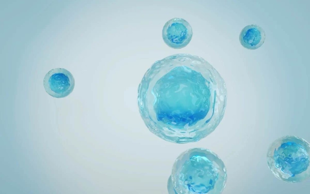
After encountering an antigen, activated B cells can differentiate into plasma cells, which are terminally differentiated B cells specialized for secreting large quantities of antibody. Other B cells can become memory B cells, which are long-lived cells that can respond rapidly when they encounter the antigen again.
Both populations represent potential sources of antibody sequences, but clone identification requires linking antibody function to the corresponding genetic sequence.
Several established technologies approach this challenge differently.
Hybridoma technology fuses an antibody-producing B cell with an immortal myeloma cell, creating a stable antibody-producing cell line. The approach preserves the heavy- and light-chain combination present in the original B cell, but cell fusion, clone expansion, and subsequent screening can limit throughput.
Antibody phage display instead presents antibody fragments on bacteriophage particles and enables very large libraries to be screened through repeated selection. In conventional combinatorial immune libraries, however, heavy- and light-chain variable regions are generally amplified in bulk and recombined during library construction. Consequently, the resulting VH/VL combinations do not necessarily represent the original pairings found within individual B cells. Specialized approaches can preserve pairing, so this is not an inherent limitation of every display workflow.
Single B cell screening bypasses the need for cell fusion by enabling direct analysis of individual B cells. Antibody genes can then be recovered from selected cells while retaining the association between their naturally paired heavy and light chains. Miniaturization, microfluidics, and sequencing are important developments for increasing the number of cells that can be examined.
Droplet microfluidics provides one way to scale this single-cell approach further.
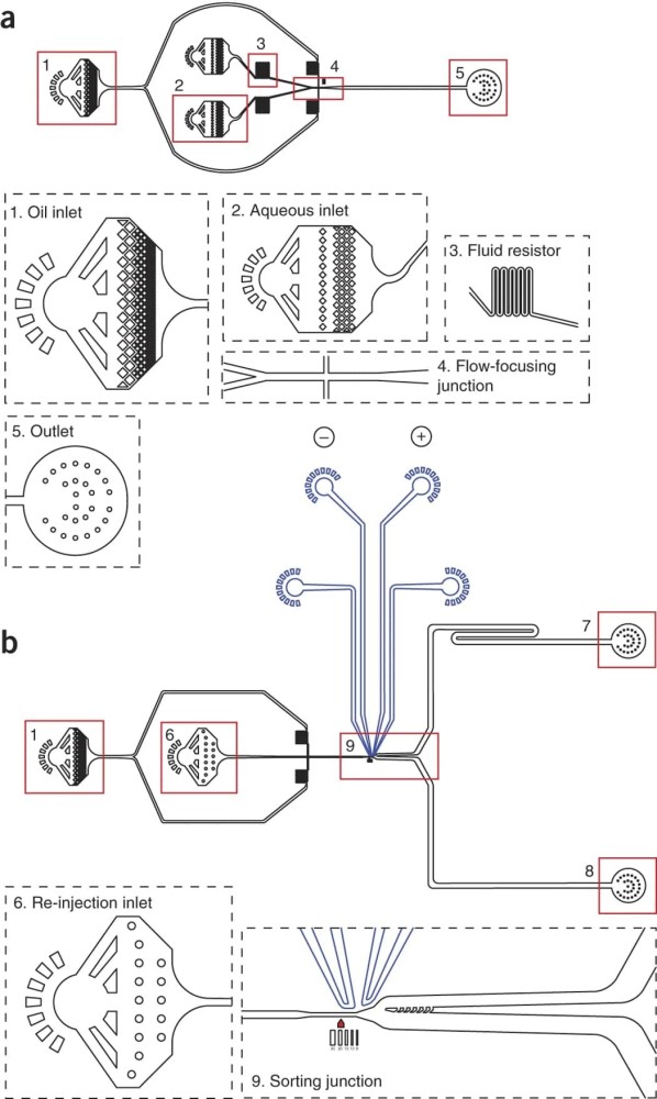
Microfluidics refers to technologies that precisely manipulate very small volumes of fluids through microscopic channels. In droplet microfluidics, one liquid is divided into discrete droplets within another immiscible liquid, which is typically an aqueous droplet surrounded by an oil phase.
The resulting droplets can have volumes in the picoliter range. A picoliter is one trillionth of a liter.
For antibody screening, cells can be diluted so that individual cells become encapsulated inside separate droplets along with the reagents required for an assay. Each droplet then functions as an isolated miniature reaction vessel.
This physical isolation is particularly useful when studying secreted molecules.
In a typical flow cytometry experiment, researchers can readily measure molecules located on or inside a cell. Measuring a protein that has already been secreted is more difficult because the molecule can diffuse away from the cell that produced it.
Droplet compartmentalization changes this. When an antibody-secreting cell is enclosed inside a droplet, secreted antibody remains confined within the same small volume. This allows an assay to measure antibody production while maintaining its association with the originating cell.
The small volume of a droplet can allow molecules secreted from an individual cell to reach detectable concentrations relatively quickly.
In an early antibody-screening example, Mazutis and colleagues encapsulated single hybridoma cells with antibody-capture beads and a fluorescent probe in approximately 50-picoliter droplets.2 Secreted antibodies could be detected after 15 minutes, and fluorescent droplets could subsequently be sorted.
The exact droplet volume, incubation time and detection strategy vary between platforms, but the principle remains the same: compartmentalization keeps the secreted antibody physically associated with the cell that produced it and concentrates the assay within a small reaction volume.
Miniaturization also reduces the amount of reagent required for each individual screening reaction, although total reagent use and cost depend on the design and scale of the complete workflow.
Producing millions of droplets does not mean that every droplet contains exactly one cell.
Cell encapsulation is typically governed by statistical loading. At concentrations chosen for single-cell experiments, some droplets will contain no cells, some will contain one cell and a smaller proportion may contain multiple cells.
Conditions must be optimized to generate enough single-cell droplets while limiting doublets, droplets containing two cells, because a doublet can make it difficult to determine which cell produced the measured signal.
Cell concentration, cell aggregation, sample quality, droplet size and the behavior of the specific cell population can all affect encapsulation.
In the published AbDrop proof-of-concept study, approximately two million droplets were generated while processing approximately one million plasma cells. Around 30-40% of droplets contained a single plasma cell, consistent with the expected statistical behavior of cell loading.3
Once a cell has been encapsulated, the next step is converting antibody secretion or antigen recognition into a measurable signal.
Different droplet platforms solve this in different ways.
Mazutis and colleagues used antibody-capture beads together with a fluorescent antigen probe. Antibody secreted by a hybridoma cell was captured on a bead, and antigen binding generated localized fluorescence that could be detected during droplet sorting.2
Shembekar and colleagues later demonstrated a droplet assay in which antibody-producing hybridoma cells were co-encapsulated with target cells. Antibodies binding the surface of those target cells produced a fluorescence signal that could be used to enrich specific antibody-producing clones.4
Ding and colleagues used a bead-based binding assay to isolate rare primary B cells producing antigen-binding antibodies. After sorting, paired heavy- and light-chain sequences were recovered from individual cells and selected antibodies were subsequently produced for confirmation by ELISA.5
Once droplets have developed a measurable signal, positive droplets can be separated from the rest of the population.
One approach is fluorescence-activated droplet sorting (FADS). FADS applies a principle similar to fluorescence-activated cell sorting, or FACS, but sorts droplets rather than individual cells in suspension.
FACS is a flow cytometry technique in which cells pass individually through a detector and are physically separated according to fluorescence or other measurable properties.
In FADS, droplets pass through an optical detection region. Fluorescence signals are measured, and droplets meeting predetermined selection criteria can be diverted into a separate collection channel.
Because droplets can be generated and interrogated rapidly, this approach can examine much larger populations than would be practical if every cell required an individual microtiter well.
Throughput is valuable when the desired antibody-producing population is rare, but screening more cells does not by itself guarantee better antibodies. Immunization strategy, antigen quality, assay design, detection thresholds and downstream validation all influence the quality of the candidates.
Selecting a positive cell is only useful for antibody discovery if its antibody sequence can subsequently be recovered.
An IgG antibody contains two identical heavy chains and two identical light chains. The variable regions of the heavy and light chains come together to form the antigen-binding surface.
The particular VH/VL combination therefore contributes directly to antibody specificity and affinity.
Single-cell sequencing methods aim to preserve the relationship between the heavy- and light-chain sequences originating from the same cell. This is often referred to as native, natural or cognate heavy/light-chain pairing.
After positive cells are recovered, their RNA can be converted into complementary DNA, or cDNA, and immunoglobulin variable-region sequences can be amplified. Modern approaches can add cell-specific molecular barcodes so that heavy- and light-chain sequences can later be assigned to the same cell during sequencing analysis.
Next-generation sequencing (NGS), massively parallel sequencing of many DNA molecules, can then be used to analyze the recovered antibody repertoire at scale.
Once antibody sequences have been identified, selected VH and VL sequences can be cloned into expression constructs and produced as recombinant antibodies.
Recombinant antibody expression means producing an antibody from a defined DNA sequence in a laboratory expression system, commonly using mammalian cells for full-length IgG production.
This step allows antibodies identified from different single cells to be produced under standardized experimental conditions and compared directly.
Downstream assays can then examine properties such as:
Antigen-specific binding
Binding affinity
Recognition of a target on the cell surface
Epitope specificity
Biological activity
Developability-related properties
Biointron's AbDrop platform applies droplet microfluidics to high-throughput plasma cell screening and integrates the screening step with antibody sequencing, bioinformatic analysis, recombinant expression and downstream validation.
In the published AbDrop workflow, plasma cells are isolated and encapsulated with fluorescently labeled antigen, fluorescent antibody-detection reagents and components supporting short-term cell culture. Following incubation, droplets generating antigen-associated fluorescence signals are selected and recovered for single-cell library preparation and sequencing.
The platform can process approximately 1-2 million plasma cells per run. Sequence analysis can recover hundreds to thousands of unique antibody sequences, with naturally paired heavy and light chains, within approximately one week. Selected antibodies can then move into high-throughput full-length IgG expression and experimental validation.
In a proof-of-concept study targeting programmed cell death protein 1 (PD-1, an immune checkpoint receptor that regulates T-cell activity), approximately one million plasma cells were processed. The study ultimately identified 461 unique IgG sequences. Fifty-two candidates were selected for antigen-binding validation, and further characterization identified antibodies with different binding and functional properties, including blocking and agonistic activity.
Droplet-based single B cell screening is one of several approaches to antibody discovery.
Hybridoma technology remains useful when stable antibody-producing clones are desirable and preserves the antibody pair generated by the original B cell.
Phage display can access very large libraries and provides considerable flexibility for antibody selection and engineering.
FACS-based single B cell approaches allow individual antigen-reactive B cells to be isolated directly based on cell-surface markers and antigen-binding probes.
Droplet-based single B cell screening is particularly suited to situations where researchers want to look at secreted antibody from individual cells while maintaining the association between the secretion phenotype and the cell's genetic information.
Several factors can influence the interpretation of a droplet-based screen.
Primary antibody-secreting cells have finite viability outside their native biological environment. Sample preparation, isolation procedures, and incubation conditions need to preserve cells long enough to produce a measurable antibody signal.
Empty droplets reduce screening efficiency, while droplets containing multiple cells can complicate the link between phenotype and genotype. Cell concentration and aggregation therefore need to be controlled.
Fluorescent labeling can potentially change an antigen or introduce assay background. Detection reagents also need sufficient specificity to distinguish true antigen recognition from nonspecific fluorescence.
Setting a selection threshold too low can increase the number of false-positive droplets, whereas a threshold that is too stringent could exclude cells producing weaker but potentially relevant signals.
Following cell selection, the corresponding heavy- and light-chain sequences must be accurately recovered. Single-cell library preparation, amplification, and sequencing are therefore integral components of the screening workflow and directly influence the quality of downstream antibody identification.
Screening hits should be confirmed using recombinantly expressed antibodies before conclusions are drawn regarding specificity, affinity, or biological activity. Validation with an independent assay format can help distinguish true binding or functional activity from assay-specific artifacts.
Droplet microfluidics enables high-throughput analysis of individual antibody-producing cells while preserving the link between antibody phenotype and the corresponding cell of origin.
By compartmentalizing cells into picoliter-scale reaction environments, secreted antibodies can accumulate around their source and be interrogated using appropriately designed detection assays. Positive droplets can then be isolated, antibody heavy- and light-chain sequences recovered, and selected sequences moved into recombinant expression and experimental validation.
Beyond increasing screening throughput, droplet-based single B cell screening enables antibody phenotype to be linked with the corresponding cell and antibody sequence across large immune repertoires.
Biointron's AbDrop Single B Cell Screening platform integrates this principle into a workflow spanning plasma cell screening, naturally paired VH/VL sequence recovery, high-throughput recombinant antibody expression and downstream validation. The approach enables up to approximately 1-2 million plasma cells to be interrogated per run, providing a route from diverse immune repertoires to experimentally characterized antibody candidates.
Cooper M. D. (2015). The early history of B cells. Nature reviews. Immunology, 15(3), 191–197. https://doi.org/10.1038/nri3801
Mazutis, L., Gilbert, J., Ung, W. L., Weitz, D. A., Griffiths, A. D, & Heyman, J. A. (2013). Single-cell analysis and sorting using droplet-based microfluidics. Nature Protocols, 8(5), 870–891. https://doi.org/10.1038/nprot.2013.046
Yu, M., Wang, L., Yang, Y., Liang, S., Ge, Y., Xi, H., Lin, S., Chen, Y., Liu, W., Wang, W., Changchun Zha, & Lu, H. (2025, December 4). AbDrop-a scalable microfluidics-enabled platform for rapid discovery and functional analysis of plasma cell-derived antibodies. mAbs; Taylor & Francis. https://www.tandfonline.com/doi/full/10.1080/19420862.2025.2597610
Shembekar, N., Hu, H., Eustace, D., & Merten, C. A. (2018). Single-Cell Droplet Microfluidic Screening for Antibodies Specifically Binding to Target Cells. Cell Reports, 22(8), 2206-2215. https://doi.org/10.1016/j.celrep.2018.01.071
Ding, R., Hung, K. C., Mitra, A., Ung, L. W., Lightwood, D., Tu, R., Starkie, D., Cai, L., Mazutis, L., Chong, S., Weitz, D. A., & Heyman, J. A. (2020). Rapid isolation of antigen-specific B-cells using droplet microfluidics. RSC advances, 10(45), 27006–27013. https://doi.org/10.1039/d0ra04328a
Biointron’s Antibodies After Hours: From Algorithms to Antibodies was held at Th……
Discover how high-throughput wet-lab validation connects AI-designed antibody se……
Understand the hybridoma generation process for monoclonal antibody production, ……
Artificial intelligence (AI) is changing how therapeutic antibodies are discover……