


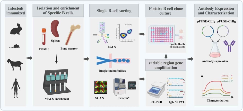


Identifying antibodies that bind a specific target is a critical step in antibody discovery. However, among the millions of B cells generated during an immune response, only a fraction may produce antibodies with the desired specificity. Distinguishing these candidates from nonspecific binders requires sensitive screening methods and reliable detection signals.
Direct antigen labeling is one approach that makes these interactions easier to detect. By attaching a fluorescent or other detectable label to a target antigen, researchers can identify B cells or secreted antibodies that recognize the target. This strategy is widely used in fluorescence-based cell sorting and can also support high-throughput microfluidic antibody screening.
However, the quality of the screening result depends on more than fluorescence intensity. Antigen structure, labeling chemistry, assay design, and background controls all influence whether a detected signal represents a genuine antigen-antibody interaction.
Direct antigen labeling involves attaching a detectable marker, typically a fluorescent dye, to an antigen so that its interaction with an antibody can be monitored.
For example, an antigen can be chemically conjugated to a fluorophore and used as a probe in fluorescence-activated cell sorting (FACS). When the labeled antigen binds to a B cell receptor (BCR) on the surface of a B cell, the resulting fluorescence allows that cell to be distinguished from cells that do not recognize the antigen.
Common fluorescent labels include phycoerythrin (PE) and allophycocyanin (APC). The choice depends on factors such as instrument compatibility, signal intensity, and the number of markers being measured simultaneously.
As described by Xu et al. (2022), antigen labeling can be achieved through chemical conjugation, recombinant fluorescent fusion proteins, or biotin-streptavidin-based methods.1
The distinction between direct and indirect detection lies in how the fluorescent signal is introduced.
In a directly labeled antigen probe, the fluorophore is attached to the antigen itself. This allows the antigen-antibody interaction to be detected without an additional fluorescent detection reagent.
Indirect detection uses another molecule to generate the signal. For example, a biotinylated antigen may be detected through fluorescent streptavidin, or an unlabeled antigen-antibody complex may be recognized by a labeled secondary reagent.
Direct labeling can simplify assay design by reducing the number of detection components. It may also help limit background associated with additional reagents. However, indirect methods can provide greater flexibility and, in some assay formats, signal amplification.
| Feature | Direct fluorescent antigen labeling | Indirect antigen detection |
|---|---|---|
| Detection method | Fluorophore attached directly to antigen | Signal generated through an additional detection reagent |
| Assay complexity | Generally fewer detection components | May require additional binding or incubation steps |
| Background | Avoids some reagent-associated background | Additional detection reagents may introduce nonspecific interactions |
| Signal strength | Depends on fluorophore properties and labeling density | May benefit from amplification or multivalent detection |
| Main consideration | Labeling must preserve antigen structure and binding activity | Detection reagents require appropriate specificity controls |
Single B cell screening enables researchers to examine individual B cells and identify those associated with antibodies of interest. Fluorescent antigen probes provide a means of detecting these interactions, whether through surface BCR binding or assays that measure antibodies secreted by individual cells.
Antigen-specific B cells can represent only a small proportion of a biological sample, making their identification challenging.
In conventional antigen-specific FACS, fluorescently labeled antigens bind to surface BCRs, allowing researchers to enrich antigen-reactive B cells before sequencing or further characterization.
This approach has become particularly valuable for studying human B cell repertoires. For example, antigen-based sorting has been used in research on SARS-CoV-2 antibodies to isolate B cells from infected or vaccinated individuals for subsequent antibody discovery.
Xu et al. (2022) highlight fluorescent antigen labeling as an important component of these workflows, while noting that low-frequency antigen-specific populations and nonspecific probe binding remain significant challenges.
Importantly, not all B cell populations are equally suited to surface antigen-based sorting. Antibody-secreting cells, particularly IgG-secreting plasma cells, may express relatively little surface immunoglobulin. For these populations, screening the antibodies they secrete provides an alternative route to identifying antigen-specific candidates.
A fluorescent signal does not necessarily indicate genuine antigen recognition. Some cells may bind nonspecifically to fluorescent dyes, streptavidin, linkers, or other assay components, producing signals that resemble true binding events.
Careful assay design helps distinguish these interactions.
One approach is dual-color antigen labeling, in which the same antigen is prepared with two different fluorophores. Cells that recognize both probes are more likely to be genuine antigen binders than cells that interact with only one labeling component.
Appropriate negative controls are equally important. These may include fluorophore-only reagents, irrelevant labeled antigens, and unlabeled antigen competition assays.
Even with these precautions, initial screening results require confirmation. Antigen labeling enables candidate identification, but it does not replace downstream validation.
Fluorescent antigen detection also plays an important role in microfluidic single-cell technologies, which allow large numbers of individual antibody-secreting cells to be examined in parallel.
Rather than identifying B cells solely through their surface receptors, these systems can isolate individual cells in small compartments and assess the antibodies they release.
A study by Winters et al. (2019) demonstrated this approach using a nanofluidic screening platform.2 Researchers isolated antibody-secreting cells in individual nanopens and used a two-color fluorescent bead-based assay to detect both IgG secretion and antigen-specific binding.
Of the 201 IgG-secreting nanopens identified, 51 also displayed antigen-specific signals. Following cell recovery and antibody sequence analysis, the researchers identified 13 unique antibody sequences.
The study demonstrates how fluorescence-based detection can connect antibody secretion, antigen recognition, and sequence recovery within a single-cell discovery workflow.
The effectiveness of antigen labeling depends on whether the labeled molecule retains the biological properties required for antibody recognition.
Poorly optimized labeling can alter protein structure, interfere with epitopes, or generate nonspecific signals that complicate candidate selection.
Antibodies recognize specific molecular features, or epitopes, on their target antigens. Many of these epitopes depend on the antigen's three-dimensional structure.
Chemical modification during labeling can interfere with recognition if a fluorescent dye or linker attaches near an important binding site. Excessive labeling may also affect protein folding, stability, or solubility.
These concerns are particularly relevant for conformational epitopes, where antibody recognition depends on the native arrangement of amino acids rather than a short linear sequence.
For this reason, antigen purity, structural integrity, and aggregation state should be assessed before and after labeling whenever possible. Where suitable reference antibodies are available, their binding to the labeled antigen can provide evidence that relevant epitopes remain accessible.
The labeling method should reflect both the properties of the antigen and the requirements of the screening assay.
Random chemical conjugation offers a relatively straightforward way to attach fluorescent dyes, but labeling can occur at multiple positions on the protein. Site-specific approaches may provide greater control over label placement and help preserve sensitive regions of the antigen.
The degree of labeling is another important variable. Too few fluorophores may produce weak signals, while excessive labeling can alter antigen behavior or increase background.
Fluorophore selection also requires attention to spectral overlap, particularly in multiplexed assays where several fluorescent probes are measured together.
Reliable screening requires separating antigen-specific binding from signals generated by the labeling reagents themselves.
| Potential challenge | Effect on screening | Possible mitigation |
|---|---|---|
| Epitope masking | Reduced detection of genuine binders | Optimize labeling sites and confirm binding activity |
| Excessive labeling | Altered antigen properties or increased background | Control labeling density |
| Antigen aggregation | Artificially strong or nonspecific binding signals | Assess aggregation before and after labeling |
| Fluorophore or streptavidin binding | False-positive events | Include reagent-only and irrelevant-probe controls |
| Spectral overlap | Difficulty distinguishing signals in multiplex assays | Select compatible fluorophores and apply appropriate compensation |
| Weak antigen-binding signals | Potential loss of low-frequency binders | Optimize probe concentration and detection conditions |
When combined with single-cell technologies, antigen labeling helps connect antibody specificity with the B cell responsible for producing that antibody.
Modern antibody discovery workflows increasingly combine single-cell screening, antibody sequencing, recombinant expression, and functional validation. This integration allows researchers to move from an initial binding signal to a characterized antibody candidate while preserving the naturally paired heavy- and light-chain sequences.
For example, droplet-based microfluidic platforms isolate individual antibody-secreting cells in miniature reaction compartments. Within each droplet, secreted antibodies can interact with labeled antigens and detection reagents. Fluorescence-based readouts then help identify droplets containing cells that produce antigen-binding antibodies.
Biointron's AbDrop Single B Cell Screening platform applies this approach to high-throughput antibody discovery. The platform uses microfluidic droplets to isolate plasma B cells and detect antigen-specific antibody secretion, followed by recovery of naturally paired antibody sequences through single-cell sequencing.
AbDrop can screen approximately 1-2 million plasma B cells in a day. Selected antibody sequences can then be advanced to recombinant expression and validation using methods such as ELISA, surface plasmon resonance (SPR), and flow cytometry.
This integrated workflow connects early antigen-binding detection with downstream antibody characterization, supporting the identification of candidates from diverse B cell populations.
AbDrop™: Microfluidic-Based Single B Cell Screening Platform →
Direct antigen labeling is a valuable tool in antibody discovery, providing a way to visualize antigen recognition and identify promising antibody-producing cells. Its applications range from conventional antigen-specific B cell sorting to fluorescence-based microfluidic screening.
However, reliable results depend on more than introducing a fluorescent label. Preserving antigen structure, selecting appropriate labeling chemistry, and controlling nonspecific interactions are essential for interpreting binding signals accurately.
As single B cell technologies continue to advance, integrating reliable antigen detection with high-throughput screening and sequence recovery will remain important for efficient antibody discovery.
Biointron's Single B Cell Screening service combines microfluidic antibody screening, antibody sequencing, and downstream validation to support the discovery of antigen-specific antibody candidates.
References:
Xu, Z., et al. (2022). Advances in antibody discovery from human BCR repertoires. Frontiers in Bioinformatics, 2, 1044975. https://doi.org/10.3389/fbinf.2022.1044975
Winters, A., et al. (2019). Rapid single B cell antibody discovery using nanopens and structured light. mAbs, 11(6), 1025–1035. https://doi.org/10.1080/19420862.2019.1624126
Biointron announces limited-time RushData launch pricing for high-throughput ant……
August 2026 brought another mix of acquisitions, licensing agreements, strategic……
Antibody discovery often begins with a highly diverse population of B cells, onl……