Antibody discovery has traditionally relied on methods like hybridoma technology and phage display, but these approaches face increasing limitations, including low throughput, disrupted antibody chain pairing, and time-consuming workflows. Microfluidics is helping to overcome these barriers by enabling faster, more efficient, and higher-resolution screening of antibody-producing cells. Across recent studies, microfluidics has shown clear advantages in accessing rare B cell subsets, preserving native antibody pairing, and integrating with sequencing and functional assays.
Droplet microfluidics is one of the most widely adopted approaches for high-throughput antibody screening. In this system, single cells are encapsulated in picoliter-scale water-in-oil droplets, which act as individual reaction chambers. A recent review showed how this method enables millions of functional antibody assays to be performed rapidly while preserving the natural pairing of heavy and light chains, which is something that is often lost in phage or yeast display systems. The droplets can be used for a range of assays, including binding, internalization, and neutralization, and can be sorted based on fluorescence signals to isolate rare antibody-producing cells. This setup also allows integration with PCR and next-generation sequencing, providing detailed information on antibody sequences linked to their function.

Plasma cells are a rich source of high-affinity, mature antibodies, but they are difficult to capture in large numbers using traditional methods. The AbDrop platform, described by Yu et al., combines droplet microfluidics with optimized workflows for isolating plasma cells at scale. The system processes up to two million plasma cells in a single run and enables recovery of hundreds to thousands of unique antibody sequences within one week. Selected clones can then be rapidly expressed and tested for antigen binding and functional activity. In a case study targeting PD-1, AbDrop successfully identified both blocking and agonist antibodies. Unlike other platforms that express antibodies as scFvs, AbDrop directly produces full-length IgGs, allowing immediate evaluation of developability and function.

Meanwhile, researchers from the University of Cambridge developed a method that uses agarose hydrogel beads to capture antibodies secreted by individual antibody-secreting cells (ASCs). Each cell is encapsulated in a microgel that immobilizes its secreted antibodies, allowing standard flow cytometry to be used for sorting based on antigen binding. This makes it possible to screen millions of cells rapidly without specialized microfluidic droplet sorters. The method was used to isolate potent, neutralizing antibodies against SARS-CoV-2 from both mouse and human ASCs within two weeks. This approach is particularly useful for studying ASCs, which typically lack surface immunoglobulin and are not accessible using traditional FACS-based antigen bait strategies.

Rabbit monoclonal antibodies are known for their high affinity and diverse epitope recognition, but they have been underutilized due to challenges in isolating rabbit B cells. A recent paper addressed this by combining magnetic enrichment with droplet microfluidics, using a customized antibody cocktail to isolate rabbit B cells without relying on standard surface markers. Two complementary droplet-based assays (one for soluble antigens and one for membrane-bound targets) were used to detect antigen-specific antibody secretion. The system demonstrated efficient recovery of high-affinity rabbit antibodies and represents a valuable expansion of species options for antibody discovery.

Another newly developed system, described by Wagner et al., uses ribosome display in combination with single-cell RNA sequencing and microfluidics to map the binding specificities of thousands of antibodies at once. Ribosome-displayed antibody fragments are incubated with mammalian cells expressing antigens on their surface, and the entire mixture is processed in droplets to link antibody and antigen information at the single-cell level. This one-step system allows profiling of antibody specificity without the need for purified, barcoded antigens. The platform was used to characterize responses to SARS-CoV-2 variants and showed how combinations of antibodies with complementary binding profiles could be selected for broader neutralization.

In addition to sequence and binding analysis, structural information about antibody-antigen interactions is increasingly important, particularly for vaccine design. Researchers from Scripps Research Institute developed a method called mEM (microfluidic electron microscopy), which enables structural epitope mapping using as little as 4 μL of serum. The system combines microfluidics with electron microscopy sample preparation and analysis, reducing processing time from over a week to less than two days. It was used to map polyclonal antibody responses to multiple viral glycoproteins, including SARS-CoV-2, influenza, and HIV Env. By providing high-resolution structural insights from small sample volumes, mEM supports both basic immunology and applied therapeutic development.

Microfluidic technologies are enabling a new generation of antibody discovery tools that are faster, more scalable, and more informative than traditional methods. By supporting single-cell resolution, preserving natural antibody pairing, and integrating functional and structural analysis, these systems offer powerful ways to access rare and diverse antibody repertoires.
Biointron’s Q2 2026 Antibody Industry Trends report aims to explore the events a……
Seasonal influenza causes an estimated one billion infections, 3-5 million sever……
Artificial intelligence can now help researchers analyze antibody sequences, pre……
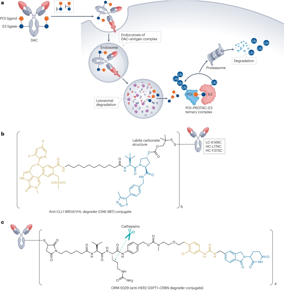
Degrader-antibody conjugates, or DACs, combine the targeting ability of antibodi……