Antibody labeling is an invaluable tool for clinical and research experiments, allowing scientists to detect and visualize the location of specific antigens in biological samples such as cells, tissues, or proteins. It is often used in conjunction with an immunoassay, and both primary and secondary antibodies can be labeled, depending on research needs.
The primary consideration when antibody labeling is retaining the antibody's activity. Generally, polyclonal antibodies (pAbs) exhibit greater versatility and resistance against activity loss compared to monoclonal antibodies (mAbs), but it is always necessary to be careful when labeling antibodies to ensure the creation of a high-quality conjugate. This is particularly true when incorporating multiple labels on the same immunoglobulin.1
There are various types of labels, including enzymes, fluorophores, biotin, colored latex beads, gold nanoparticles, and metal tags. The most popular sites on antibodies to label or cross-link biomolecules are the charged amino acids found on the outer surface of the antibodies. There are four main targets for labeling antibodies, the other three being carbohydrate moieties, sulfhydryl groups found on cysteine residues, and tyrosine residues.
The amine (NH2) groups of antibodies are abundantly present in lysine residues and at the N-termini of polypeptide chains, enabling labeling at multiple sites. They are easily modified due to their reactivity, and carboxyl groups on the side chains of glutamic acid and aspartic acid may be utilized for cross-linking.
Carbohydrate moieties are often found on the highly glycosylated antibody Fc region. Labeling in this region is beneficial since they are far from the antigen recognition site, so there is a decreased risk of interference. In addition, if oxidized to aldehydes, the antibody’s antigen-binding affinities will not be significantly affected.
Labeling at sulfhydryl-bearing cysteines is another option, as reduction of cysteines will divide the antibody into two monovalent domains without damaging antigen-binding sites. Sulfhydryls are formed when cysteine is oxidized to form a disulfide bond, which is then further reduced at the hinge region of the antibody.
Another alternative labeling target is at tyrosine residues, which can be iodinated to radiolabel the antibody. The United States FDA has approved 90Y-a pure β-emitter, and I131-a dual β and γ emitter, as therapeutic radionuclides for conjugating antibodies for cancer therapy, in addition to 111In and 99mTc (γ emitters) labeled mAbs for diagnostic uses.2
Berg, E. A., & Fishman, J. B. (2020). Labeling Antibodies. Cold Spring Harbor protocols, 2020(7), 099242. https://cshprotocols.cshlp.org/content/2020/7/pdb.top099242
Gupta, S., Batra, S., & Jain, M. (2014). Antibody Labeling with Radioiodine and Radiometals. Methods in Molecular Biology (Clifton, N.J.), 1141, 147. https://link.springer.com/protocol/10.1007/978-1-4939-0363-4_9
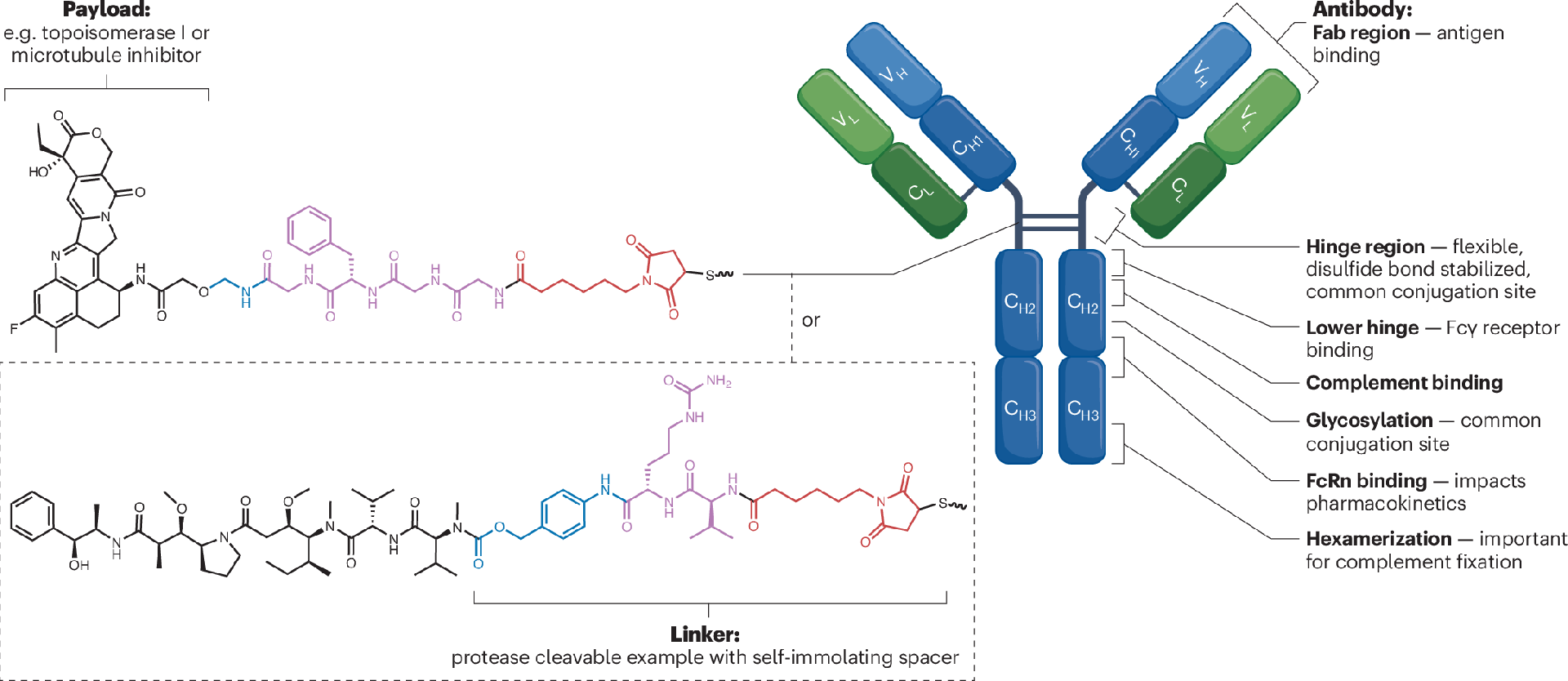
In this article, we explore the major trends shaping the future of targeted canc……
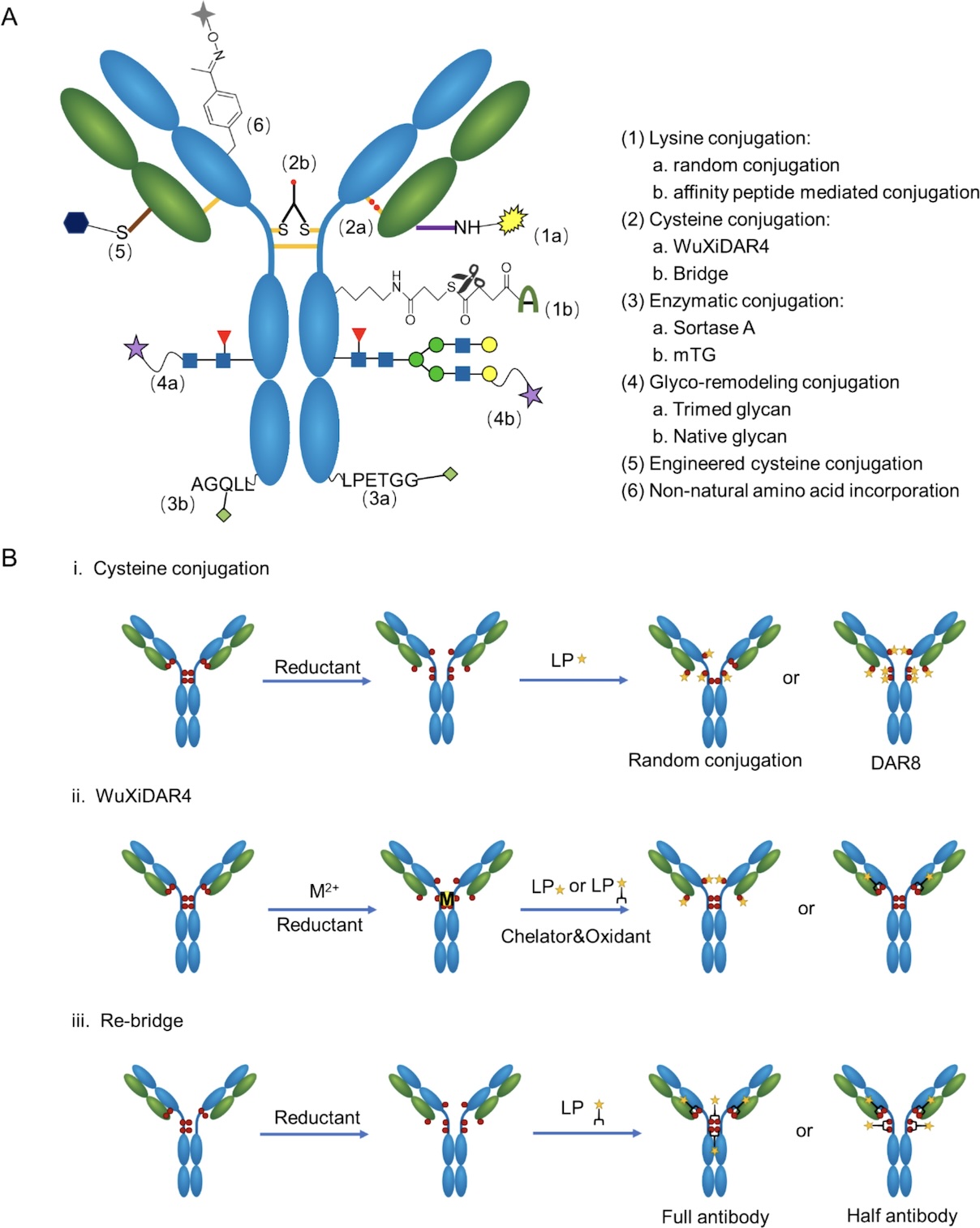
Site-specific conjugation limits payload attachment to defined positions on the ……
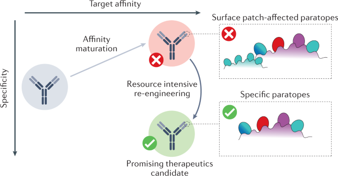
Affinity maturation can improve binding to the intended target, but it can also ……
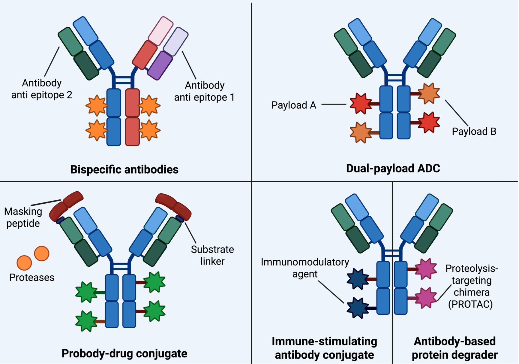
ADC development is no longer limited to the antibody-linker-toxin model. The fie……
You can also contact us on the Scientist and Science Exchange marketplaces.
Our website uses cookies to improve your experience. Read our Privacy Policy to find out more.
Biointron uses cookies and similar technologies to ensure proper website functionality, understand how visitors use our website, and improve our content and services. Necessary cookies are enabled by default.
By clicking “Accept All”, you agree to the use of all cookies, including analytics and optional cookies that help us understand website usage and improve user experience. By clicking “Reject Non-Essential”, only necessary cookies will be used.
You may manage or change your cookie preferences at any time by clicking “Cookie Settings” at the bottom of the website.
For more information about how we use cookies and process personal data, please review our Privacy Policy.