




Fluorescent probes are molecular tools that use fluorescent molecules (fluorophores) to generate detectable signals when excited by light at an appropriate wavelength. They typically consist of a targeting component, such as an antibody, VHH, peptide, nucleic acid, or small molecule, linked to a fluorophore. When the targeting component binds to its specific target, the fluorescent signal can be used to visualize or quantify molecular targets and cellular processes using techniques such as fluorescence microscopy and flow cytometry.
Fluorescent probes are widely used in biological research, diagnostics, and molecular imaging. Applications include tracking protein localization and cellular processes, detecting biomarkers, and visualizing tumors during fluorescence-guided surgery. Fluorophores differ in properties such as brightness, excitation and emission wavelengths, photostability, and susceptibility to environmental conditions. The choice of fluorophore should therefore be matched to the intended application and imaging platform, such as confocal microscopy, super-resolution microscopy, or two-photon imaging.
When conjugated to high-affinity targeting molecules such as VHHs, fluorescent probes can provide selective detection of proteins and other molecular targets in complex biological samples. The small size of VHHs can also be advantageous for certain imaging applications where probe size and tissue penetration are important considerations.
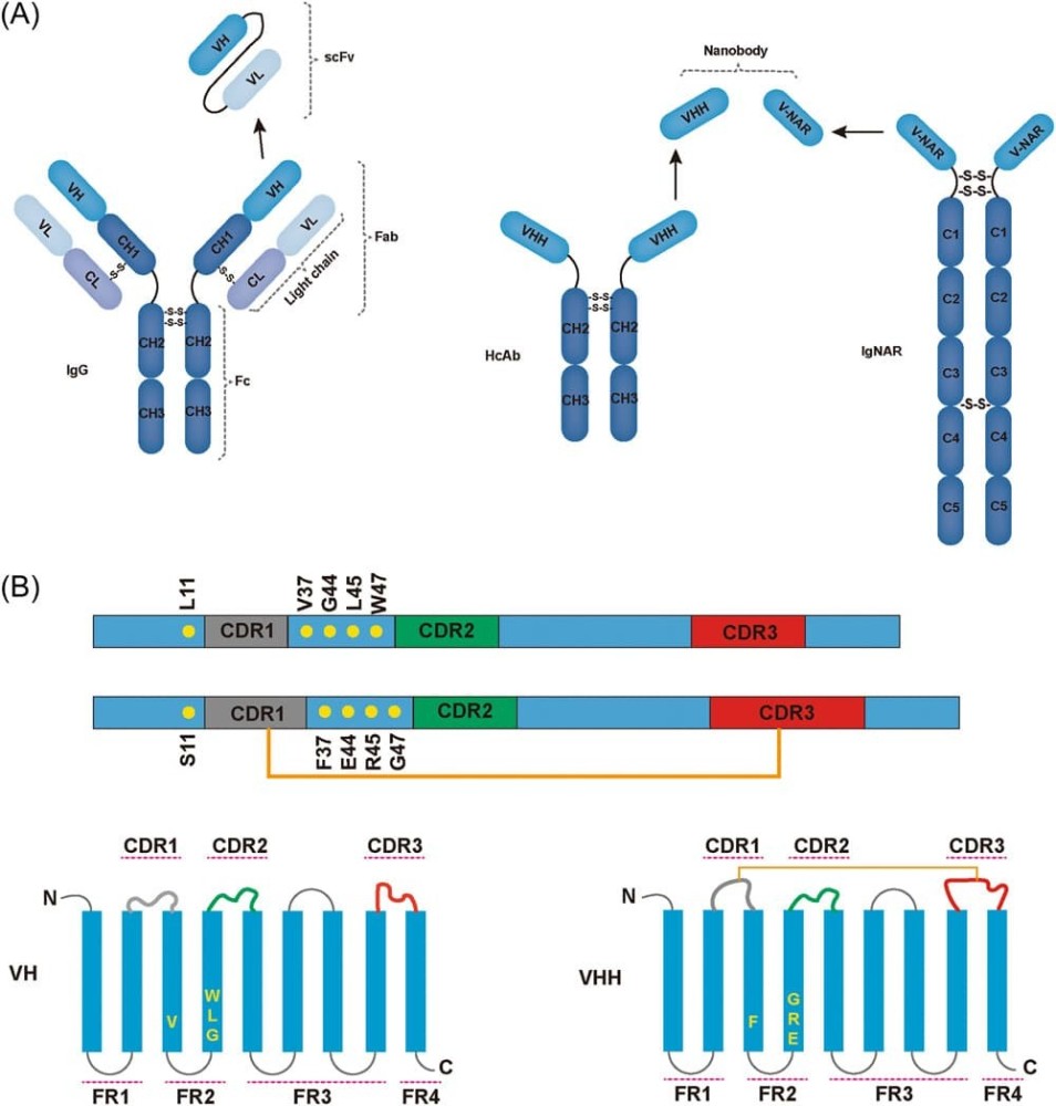
VHH antibodies, also known as nanobodies, are single-domain antigen-binding fragments derived from the heavy-chain-only antibodies found naturally in camelids. Unlike conventional monoclonal antibodies (~150 kDa), which consist of two heavy chains and two light chains, VHHs comprise a single variable heavy-chain domain. With a molecular weight of approximately 15 kDa and a compact structure of roughly 4 × 2.5 × 3 nm, VHHs can access epitopes that may be sterically difficult for conventional antibodies to reach. Their single-domain architecture can also facilitate recombinant expression and engineering.
Structurally, VHHs retain the characteristic immunoglobulin fold but often feature relatively long CDR3 loops that can contribute to recognition of recessed or conformationally constrained epitopes. Some VHHs also contain additional disulfide bonds, including those connecting CDR1 and CDR3, which can further stabilize the domain. In addition, characteristic amino acid substitutions in framework region 2 (FR2), including substitutions such as V37F and G44E, help reduce the hydrophobic surface that would normally interact with a light chain and can contribute to the solubility of VHHs as isolated domains. However, these structural features vary among individual VHHs and are not universal.
Many VHHs are identified through library-based selection approaches such as phage display, which can enable the screening and enrichment of antigen-specific binders. Subsequent engineering and screening can be used to optimize properties such as affinity, specificity, stability, and expression for particular applications.
Researchers seeking customized VHH formats and optimization strategies can use antibody engineering services to develop VHHs for imaging and diagnostic applications. Their small size and modular architecture make VHHs attractive recognition elements for fluorescent probes and other molecular imaging tools. VHHs can be expressed recombinantly in microbial or other suitable expression systems and purified using affinity tags such as His or FLAG. Site-specific labeling can be achieved through approaches including engineered cysteine residues for thiol-reactive fluorophore conjugation or enzymatic labeling strategies such as AviTag-mediated biotinylation, depending on the intended application. Fluorophores can then be selected based on properties such as brightness, photostability, excitation and emission spectra, and compatibility with the imaging platform.
These features allow VHHs to serve as antigen-specific recognition elements in fluorescent imaging probes, biosensors, and diagnostic assays. Their small size, binding specificity, and potential for recombinant engineering make them useful across a range of research and diagnostic applications. Recent reviews of nanobody applications in diagnostics highlight their potential across multiple detection and diagnostic platforms.
Fluorescently labeled VHHs are used in both live-cell and fixed-cell imaging. Their compact size can reduce the distance between the target epitope and fluorescent label, minimizing linkage error and supporting more precise localization in high-resolution imaging. Actual localization accuracy, however, depends on factors such as probe geometry, labeling strategy, and epitope position.
Two examples of VHH-based fluorescent probe formats are:
Chromobodies: VHHs genetically fused to fluorescent proteins for live-cell visualization of endogenous or tagged proteins.
Fluobodies: VHHs conjugated to fluorescent dyes for use as purified imaging probes in fixed- or, with appropriate delivery, live-cell applications.
VHH-based probes have been used to study protein localization and dynamics in mammalian cells and model organisms, including Drosophila, Zebrafish, and C. elegans. They have also been incorporated into genetically encoded biosensors for applications such as intracellular calcium and pH imaging.
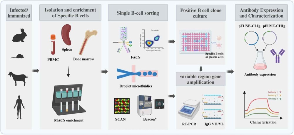
Engineered fluorescent VHHs can further optimize target specificity and, in some applications, improve signal-to-background ratios. Development may begin with approaches such as single B cell antibody discovery, followed by VHH engineering and fluorescent labeling to optimize affinity, stability, and imaging performance. For deeper research, see nanobody probes in microscopy.
One example of an emerging approach in VHH probe engineering is the Quenchbody, a fluorescent VHH designed for signal-on detection of antigen binding. Quenchbodies are typically labeled with a fluorophore positioned near tryptophan residues in the VHH. In the unbound state, interactions between the fluorophore and nearby aromatic residues can suppress fluorescence. Antigen binding alters the local environment of the fluorophore, resulting in increased fluorescence and a measurable signal.
This format enables homogeneous, mix-and-read antigen detection without the washing steps required in many conventional immunoassays. In the cited study, molecular dynamics simulations and mutational analysis were used to optimize tryptophan positioning and improve fluorescence response.
For example, the reported IL-6-binding Quenchbody produced approximately a 2.4-fold fluorescence increase upon antigen binding, with reported EC₅₀ values of 7–14 nM. This represented an improvement over earlier Quenchbody designs evaluated with other targets, although performance depends on the VHH sequence, fluorophore, target, and probe configuration.
VHH probes can also be engineered with functional tags for downstream applications, including:
Cys-tags for site-specific fluorophore conjugation
AviTags for site-specific biotinylation
FLAG tags for purification or detection
Depending on the construct and production method, engineered VHH probes can be generated using recombinant or cell-free expression systems and subsequently characterized for binding and fluorescence response.
VHH-based fluorescent probes and other VHH formats have been investigated across a range of diagnostic and biosensing platforms:
ELISA and fluorescent immunoassays: VHHs can provide specific target recognition and stable recombinant reagents for assay development.
Lateral flow assays: Their small size and recombinant production can support compact, stable detection formats.
Electrochemical biosensors: VHHs can be immobilized on sensor surfaces for selective target recognition.
VHHs can also offer favorable stability under challenging physicochemical conditions, although stability varies among individual sequences and should be experimentally validated for each application. Their small size may support tissue and cellular access in certain imaging and in vivo applications, while their relatively short systemic half-life can be advantageous for some imaging applications.
These properties, together with their amenability to recombinant engineering and site-specific conjugation, make VHHs promising recognition elements for laboratory-based diagnostics, biosensors, and point-of-care assays. For therapeutic and translational applications, antibody humanization can be used to reduce potential immunogenicity while maintaining target-binding properties.
Fluorescent VHH probes are being investigated for intraoperative tumor imaging, where rapid and selective visualization of malignant tissue could support image-guided surgery.
In an orthotopic pancreatic cancer model, a CEA-targeting nanobody conjugated to IRDye 800CW was administered intravenously. Tumor-associated fluorescence was detected shortly after administration, with a reported tumor-to-background ratio (TBR) of 2.66 at 3 hours post-injection. The probe also enabled visualization of tumors smaller than 3 mm, while studies using low-CEA-expressing tumors and control VHHs supported target-specific accumulation.
The probe incorporated a C-terminal cysteine for site-specific maleimide conjugation, an approach that can provide more controlled fluorophore attachment than nonspecific labeling. Compared with full-length antibodies, the smaller size of VHHs can enable faster tissue distribution and clearance, potentially allowing imaging within a shorter interval after administration. This makes VHH-based probes promising candidates for applications where rapid tumor-to-background contrast is desirable.
Near-infrared fluorophores such as IRDye 800CW are particularly useful for intraoperative imaging because their emission falls within a spectral range suitable for fluorescence-guided surgery. The compatibility of a given VHH probe with specific surgical imaging systems, however, depends on the fluorophore, imaging hardware, and regulatory status of the system.
Biointron offers comprehensive support for VHH antibody discovery and production
Custom VHH Discovery: Using immunized libraries.
High-Yield Expression: Mammalian systems with purification tags.
Quality Control: Stability testing, affinity validation, and dye compatibility checks.
Trusted by over 3,000 research teams worldwide, Biointron has supported antibody innovation for more than a decade. Explore our Antibody Discovery Services to accelerate your next VHH or antibody development project with expert support and proven workflows.
References:
Su, Q., Shi, W., Huang, X., Yin, S., Yang, X., & Lu, X. (2023). Recent advances of nanobody applications in diagnosis and detection. MedComm – Biomaterials and Applications, 2(3), e54. https://doi.org/10.1002/mba2.54
de Beer, M. A., & Giepmans, B. N. G. (2020). Nanobody-Based Probes for Subcellular Protein Identification and Visualization. Frontiers in cellular neuroscience, 14, 573278. https://doi.org/10.3389/fncel.2020.573278
Cater, J.H., El Salamouni, N.S., Mansour, G.H. et al. Optimised nanobody-based quenchbodies for enhanced protein detection. Commun Biol 8, 937 (2025). https://doi.org/10.1038/s42003-025-08359-3
Lwin, T. M., Hernot, S., Hollandsworth, H., Amirfakhri, S., Filemoni, F., Debie, P., Hoffman, R. M., & Bouvet, M. (2020). Tumor-specific near-infrared nanobody probe rapidly labels tumors in an orthotopic mouse model of pancreatic cancer. Surgery, 168(1), 85–91. https://doi.org/10.1016/j.surg.2020.02.020
Sograte-Idrissi, S., Oleksiievets, N., Isbaner, S., Eggert-Martinez, M., Enderlein, J., Tsukanov, R., & Opazo, F. (2019). Nanobody Detection of Standard Fluorescent Proteins Enables Multi-Target DNA-PAINT with High Resolution and Minimal Displacement Errors. Cells, 8(1), 48. https://doi.org/10.3390/cells8010048
Biointron announces limited-time RushData launch pricing for high-throughput ant……
August 2026 brought another mix of acquisitions, licensing agreements, strategic……
Antibody discovery often begins with a highly diverse population of B cells, onl……
As larger candidate panels move from computational design into the laboratory, a……