





Single-domain antibodies (sdAbs) or VHH antibodies, derived from camelid heavy-chain-only antibodies (HCAbs) found in camelids, are gaining traction as molecular imaging agents due to their distinct structural and biophysical features. These antibodies, known as VHHs or nanobodies, are approximately 12–15 kDa in size and maintain full antigen-binding capacity through a single variable domain. Their small size (2.5 × 4 nm), monomeric nature, and elongated CDR3 regions allow access to sterically hindered or cryptic epitopes inaccessible to conventional IgG molecules. These single-domain antibodies also demonstrate advantages over larger antibody formats in molecular imaging workflows.
Structurally, nanobodies share homology with the human VH3 germline, minimizing immunogenicity risks. Humanized VHHs and camelized VH domains are increasingly explored to further reduce clinical liabilities while maintaining binding affinity. Unlike conventional monoclonal antibodies (~150 kDa), nanobodies can be cloned and expressed as single-chain antibodies or single-domain constructs in microbial hosts, streamlining nanobody production, functional screening, and downstream characterization.
Their high refolding capacity and tolerance to harsh conditions (low pH, proteases, heat) support applications in both in vivo imaging and ex vivo diagnostics. These intrinsic properties make nanobodies excellent scaffolds for imaging probe development across modalities including PET, SPECT, fluorescence, ultrasound, and hybrid techniques. In addition, VHH fragments can be engineered into bispecific formats or fused with reporter systems for multimodal imaging applications.
The development of nanobody-based imaging probes involves three major stages: library construction, binder screening, and recombinant production. VHH library generation commonly begins with antigen immunization and immune system priming in camelids, leading to the isolation of antigen-specific B cells. Immune libraries, derived from antigen-immunized camelids (e.g., llama, alpaca), offer high-affinity binders and support downstream affinity maturation workflows. Alternatively, naïve and synthetic libraries, including synthetic Nb library platforms, enable rapid screening for targets where immunization is impractical. Selection platforms include phage display, yeast display, and bacterial surface display, all of which are compatible with high-throughput workflows and iterative biopanning rounds.
Recombinant production typically employs Escherichia coli as the expression host due to its rapid doubling time, established fermentation protocols, and low production costs. Many workflows rely on periplasmic secretion in E. coli to improve disulfide bond formation and protein folding efficiency during antibody production. For applications requiring post-translational modifications or more complex formats, Pichia pastoris, insect cells, or mammalian cells may be used. Purification often relies on affinity tags (e.g., His₆), although these may be removed post-purification to reduce immunogenicity or non-specific interactions in vivo. Analytical workflows frequently incorporate size-exclusion chromatography, liquid chromatography, and mass spectrometry to confirm purity, aggregation state, and molecular integrity.
Scalability, high yield, and retention of antigen-binding activity are critical for the success of nanobody-based imaging probes. Biointron’s VHH Antibody Discovery platform enables streamlined progression from gene to high-purity protein, supporting both early-stage screening and translational research. The platform also supports scalable production, downstream processing, and validation workflows for emerging VHH therapies.
VHH antibodies are compatible with a range of radiolabels for nuclear imaging. SPECT-based imaging typically uses γ-emitting radionuclides such as ⁹⁹ᵐTc and ¹¹¹In, while PET imaging utilizes positron-emitting isotopes like ⁶⁸Ga, ⁶⁴Cu, and ⁸⁹Zr. These workflows support both positron emission tomography and single photon emission computed tomography applications. Radiolabeling is commonly achieved through chelator conjugation (e.g., DOTA, NOTA, Df-Bz-NCS) or direct labeling using hexahistidine tags and tricarbonyl chemistry, particularly with ⁹⁹ᵐTc.
Clinical relevance has been demonstrated using anti-EGFR nanobodies (7C12, 7D12), anti-HER2 constructs (2Rs15d), and others targeting VCAM-1, MMR, and CEA. Imaging with ⁶⁸Ga-labeled nanobodies enables high-contrast PET imaging within 1–2 hours post-injection, a significant improvement over full-length IgGs which require delayed imaging due to prolonged circulation. Several imaging workflows also include elisa screening, affinity capture, and antigen validation steps to verify target specificity before in vivo studies.
In addition to radiolabeling, nanobody conjugates have also been applied in MRI. One study developed anti-HER2 VHH-conjugated magnetoliposomes (MLs) for high-resolution breast cancer imaging. These VHH-ML conjugates retained their binding capacity and demonstrated potent internalization into HER2-positive cells. Compared to Herceptin-MLs, anti-HER2 VHH-MLs produced higher contrast in HER2-positive versus HER2-negative cells, even at low cell density. This improved contrast was attributed to faster internalization kinetics and more efficient target engagement, highlighting the value of nanobody-MRI conjugates in tumor localization.
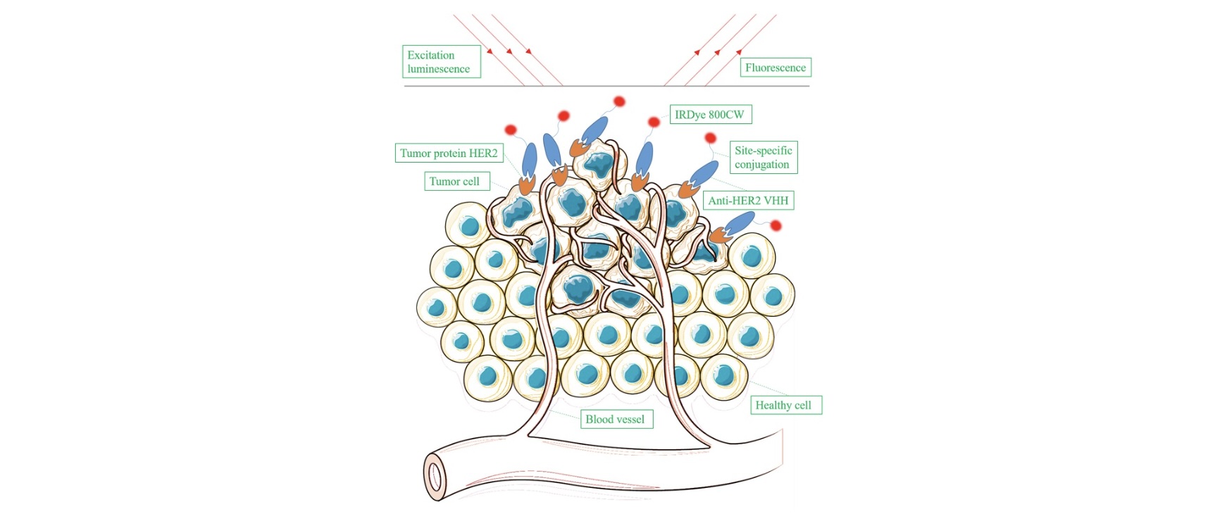
Fluorescent dyes (e.g., IRDye800CW) conjugated to nanobodies support real-time intraoperative imaging. Optical imaging with IRDye800CW-labeled anti-HER2 VHHs has shown rapid tumor accumulation and high tumor-to-background (T/B) ratios. Compared to Trastuzumab-IR, VHH-based probes accumulated ~20 times faster, enabling same-day imaging protocols and reducing delays before surgical intervention. In lymphoma and glioblastoma models, near-infrared-labeled VHHs targeting MHC-II and IGFBP7 demonstrated high specificity and signal stability for up to 96 hours post-injection. These properties support applications ranging from preoperative imaging to real-time surgical guidance. In models of HER2-positive tumors, optical imaging with dye-labeled nanobodies achieved tumor-to-background ratios exceeding those observed in SPECT/PET. Image-guided surgery applications benefit from nanobodies' rapid tumor penetration and renal clearance.
Ultrasound imaging has also been adapted for nanobody-based probes via conjugation to contrast-enhancing microbubbles. In murine models, VCAM-1-targeted microbubble probes demonstrated rapid signal acquisition and high specificity for tumor vasculature.
Related: Site-Specific Conjugation Techniques for Antibody-Based Diagnostics
Nanobody probes targeting oncogenic receptors such as EGFR, HER2, and CEA have demonstrated robust imaging performance in preclinical models. For instance, ⁶⁸Ga-2Rs15d enables PET imaging of HER2-positive tumors within 1 hour post-injection, outperforming traditional antibody tracers in both resolution and clearance kinetics.
In optical imaging, anti-HER2 nanobodies conjugated with NIR dyes have been used for successful intraoperative delineation and resection of tumor xenografts in mice. These applications are progressing to clinical trials (e.g., EudraCT 2012-001135-31), validating their translational potential.
Optical imaging also supports multiplexed tumor profiling using dual VHH probes targeting independent tumor markers. Combining VHHs conjugated to distinct fluorophores improved T/B ratios and enhanced detection of small metastases. In HER2-positive models, dual-labeled anti-HER2 VHHs not only increased contrast but also allowed intraoperative visualization of tumor margins. Site-specific conjugation at the C-terminal cysteine minimized interference with binding affinity, preserving high target specificity.
Furthermore, bimodal nanobody probes have been developed, combining radiolabels (e.g., ¹¹¹In) with fluorophores such as IRDye700DX. These agents enable dual imaging (e.g., SPECT and fluorescence) while also serving as vehicles for targeted photodynamic therapy (PDT). In one study, an anti-EGFR VHH labeled with ¹¹¹In-DTPA and IRDye700DX homed to EGFR-overexpressing tumors, enabling visualization and selective cytotoxicity upon light activation. A separate HER2-targeting VHH-IRDye700DX conjugate demonstrated complete regression of trastuzumab-resistant tumors following a single PDT session.
Radiolabeled nanobodies against macrophage mannose receptor (MMR) have been utilized to monitor tumor-associated macrophages and joint inflammation in rheumatoid arthritis models. Anti-MMR nanobodies achieved high specificity and enabled visualization of inflamed joints using SPECT/CT within 3 hours of injection.
VCAM-1-targeting nanobodies have shown promise in imaging atherosclerotic lesions in ApoE-deficient mice. Radiolabeled and microbubble-conjugated formats allow non-invasive tracking of endothelial inflammation, offering early detection potential in cardiovascular pathology.
Nanobody-based ELISA and LFIA assays have been developed for pathogens including Clostridioides difficile, Botulinum neurotoxin, SARS-CoV-2, and HEV. In SARS-CoV-2 detection, LFIA platforms using VHH conjugated to gold nanoparticles identified spike protein variants with high sensitivity.
Electrochemical biosensors incorporating nanobody receptors allow rapid, label-free antigen detection under variable conditions. These features meet WHO’s ASSURED criteria for point-of-care diagnostics, positioning VHHs as ideal tools in global health initiatives.
Small Size: Facilitates rapid tissue penetration and deep tumor access.
Fast Clearance: Enables early imaging and high tumor-to-background contrast.
Thermal and Chemical Stability: Supports robust labeling and storage conditions.
Specificity and Affinity: High target selectivity, with KD values in the low nanomolar to picomolar range.
Modality Flexibility: Compatible with PET, SPECT, optical imaging, and ultrasound.
In PET imaging, nanobody probes consistently achieve >4% injected dose per gram (ID/g) in tumor tissue with minimal non-specific uptake outside of clearance organs (e.g., kidneys). For surgical applications, rapid tumor labeling with optical probes minimizes procedural delays and enhances resection accuracy.
One of the primary challenges in nanobody imaging is renal retention. High kidney uptake can obscure lesions near the renal system and elevate organ doses. Strategies to reduce this include:
Co-injection of lysine or gelofusine, which inhibit megalin-mediated endocytosis
Removal of polyhistidine tags post-purification
Co-administration of cold (unlabeled) bivalent nanobodies to occupy off-target sites
While rapid clearance is advantageous for imaging, it may limit tumor accumulation. Half-life extension via fusion to albumin-binding domains or PEGylation has been explored but may affect tumor penetration. Applications must balance these trade-offs based on imaging time windows and disease context.
Circulation half-life remains a limiting factor for certain imaging modalities. Comparative studies have shown that bivalent (e.g., EG2-hFc, ~80 kDa) and pentavalent (~128 kDa) VHH constructs extend systemic half-life and enhance tumor accumulation. Among these, bivalent VHHs strike a balance between improved serum persistence, maintained tumor penetration, and high binding affinity. These engineered formats reduce the need for repeated dosing and are suitable for intracranial imaging applications.
Another technical consideration is the impact of fluorophore conjugation on VHH affinity. Random labeling can interfere with antigen binding, especially when dyes are conjugated near the paratope. Site-specific conjugation strategies, such as C-terminal cysteine labeling, preserve the binding activity and improve reproducibility across imaging batches.
Site-specific labeling is crucial to preserve binding affinity. Conjugation through C-terminal tags, engineered cysteines, or enzyme-mediated linkages (e.g., sortase) minimizes interference with antigen binding and allows uniform probe production.
Though native nanobodies are weakly immunogenic, certain elements (e.g., tags, linkers, bacterial residues) can provoke immune responses. Humanization and stringent purification protocols mitigate this risk, especially in clinical or repeat-use scenarios.
Advancements in imaging instrumentation have increased sensitivity and reduced noise in molecular imaging. For example:
High-resolution PET systems now allow quantification at picomolar levels
Optical imaging in the NIR window reduces background autofluorescence
Ultrasound contrast agents enhance vascular imaging with minimal invasiveness
Ongoing trials are evaluating nanobody probes in cancer staging, surgical guidance, and therapy monitoring. The combination of nanobody engineering with hybrid imaging (e.g., PET/MRI) offers further opportunities for multiplexed disease assessment.
Clinically, ⁶⁸Ga-labeled anti-HER2 VHHs have been evaluated in phase II trials to assess HER2 expression in both primary and metastatic breast cancer. These studies demonstrated favorable biodistribution, high tumor accumulation, and safety across patient cohorts. A separate phase I study assessed a ⁶⁸Ga-labeled VHH against CD206 for imaging tumor-associated macrophages, offering new options for visualizing the tumor microenvironment.
Additionally, VHHs used in imaging have shown compatibility with therapeutic antibodies. For instance, anti-CD38 nanobodies retained their target recognition even after Daratumumab binding, allowing imaging and monitoring in therapeutic contexts without competition. This suggests potential for nanobody imaging to complement monoclonal antibody therapies in clinical settings.
Nanobody-based probes represent a major step forward in molecular imaging. Their biochemical advantages (size, high affinity, fast clearance) enable rapid and high-contrast imaging across multiple modalities. Applications span oncology, inflammation, cardiovascular disease, and infectious diagnostics.
Central to this innovation is the ability to produce VHHs at scale, with high yield and consistent quality. Services such as Biointron’s VHH Antibody Discovery platform enable researchers to rapidly generate and screen high-affinity nanobodies, expediting the translational pipeline from bench to bedside.
References:
Barakat, S., Berksöz, M., Zahedimaram, P., Piepoli, S., & Erman, B. (2022). Nanobodies as molecular imaging probes. Free Radical Biology and Medicine, 182, 260-275. https://doi.org/10.1016/j.freeradbiomed.2022.02.031
Chakravarty, R., Goel, S., & Cai, W. (2014). Nanobody: The “Magic Bullet” for Molecular Imaging? Theranostics, 4(4), 386–398. https://doi.org/10.7150/thno.8006
Alexander, E., & Leong, K. W. (2024). Discovery of nanobodies: a comprehensive review of their applications and potential over the past five years. Journal of Nanobiotechnology, 22(1), 661. https://doi.org/10.1186/s12951-024-02900-y
Li, S., Hoefnagel, S. J., & Krishnadath, K. K. (2023). Single domain Camelid antibody fragments for molecular imaging and therapy of cancer. Frontiers in Oncology, 13, 1257175. https://doi.org/10.3389/fonc.2023.1257175
Cong, Y., Devoogdt, N., Lambin, P., Dubois, L. J., & Yaromina, A. (2024). Promising Diagnostic and Therapeutic Approaches Based on VHHs for Cancer Management. Cancers, 16(2), 371. https://doi.org/10.3390/cancers16020371
De Beer, M. A., & Giepmans, B. N. (2020). Nanobody-Based Probes for Subcellular Protein Identification and Visualization. Frontiers in Cellular Neuroscience, 14, 573278. https://doi.org/10.3389/fncel.2020.573278
Biointron’s Antibodies After Hours: From Algorithms to Antibodies was held at Th……
Discover how high-throughput wet-lab validation connects AI-designed antibody se……
Understand the hybridoma generation process for monoclonal antibody production, ……
Artificial intelligence (AI) is changing how therapeutic antibodies are discover……