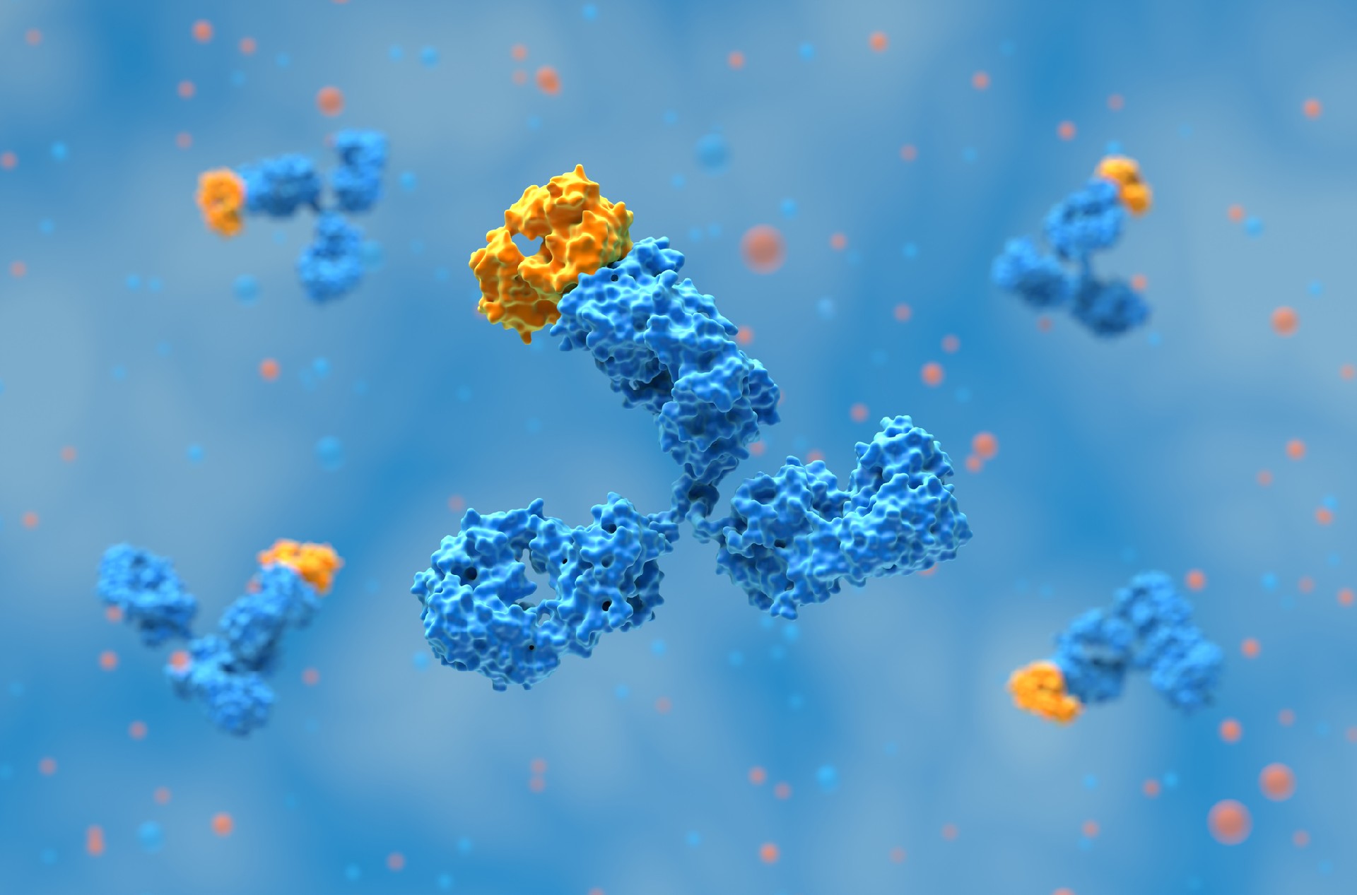
VHH antibodies, also referred to as nanobodies, are the variable domains of camelid heavy-chain-only antibodies. Their small size (~15 kDa) and high solubility make them suited for intracellular applications, and a subset of VHHs can be selected or engineered to retain binding activity when expressed in reducing intracellular compartments. These "intrabodies" can be expressed inside living cells, where they function to modulate or visualize endogenous proteins.
In contrast to conventional immunoglobulins that require oxidative environments for disulfide bond formation, engineered VHHs can retain binding activity in the reducing cytoplasmic environment. This feature, combined with their structural robustness and high binding specificity, makes them useful as genetically encoded binders for intracellular targets that are difficult to address with conventional antibody formats.
As interest in targeting intracellular protein-protein interactions grows in both therapeutic and functional genomics contexts, VHH intrabodies have emerged as precise tools for real-time cell biology and molecular medicine.
Biointron’s VHH Antibody Discovery Service offers an efficient pipeline to generate functional VHHs suitable for intracellular expression, supporting both basic research and therapeutic development pipelines.
In conventional IgG antibody formats, intradomain disulfide bonds are essential for correct folding and function. However, the cytoplasm’s reducing environment hinders disulfide bond formation. VHHs with reduced dependence on the canonical intradomain disulfide bond are preferred for cytosolic intrabody applications. While disulfide-free VHHs can be engineered through cysteine-to-serine mutations, these modifications often reduce stability and binding affinity, necessitating rigorous downstream functional screening.
Physicochemical characteristics such as charge distribution, hydrophobic surface exposure, and isoelectric point influence solubility and aggregation. Intracellularly functional VHHs are often enriched for favorable developability properties, including high solubility and limited aggregation propensity.
The extended CDR3 loops of many VHHs can engage concave surfaces, grooves, or enzyme active sites that are less accessible to conventional antibody formats. This feature expands their potential as inhibitors or biosensors targeting functional protein surfaces inside the cell.
VHH intrabodies have been applied to target intracellular aggregates of α-synuclein and tau. These nanobodies inhibit fibril formation and have been reported to interfere with α-synuclein aggregation or toxicity in selected in vitro, cellular, and preclinical model systems. For instance, α-synuclein-binding nanobodies, including NbSyn87-derived tools, have been investigated for monitoring or modulating α-synuclein species; however, there have been reported cross-reactivity and model-dependent effects.
Intracellular VHHs have been used experimentally to modulate signaling proteins, including GPCR-related pathways, although most applications remain preclinical or tool-based. One approach redirected p53 to mitochondria using a mitochondrial targeting sequence, affecting tumor cell viability.1 Other intrabodies targeting the Gβγ subunit of GPCRs were shown to disrupt downstream signaling by competing with endogenous effectors.
VHH intrabodies targeting intracellular regions of G protein-coupled receptors (GPCRs) have shown potential for modulating signaling in a therapeutically relevant context. For example, VHHs such as Nb80 have been used as conformational biosensors or stabilizing binders for active GPCR states, enabling analysis of receptor activation, trafficking, and compartmentalized signaling. These intrabodies modulate downstream pathways such as cAMP production and β-arrestin recruitment, demonstrating their utility for dissecting GPCR signaling mechanisms in live cells.
Phenotypic lentiviral screens have identified intracellularly expressed VHHs that protect cells from influenza A virus or vesicular stomatitis virus infection. Such intrabodies provide an alternative route to antiviral intervention by blocking viral replication at intracellular checkpoints.
Chromobody technology combines VHHs with fluorescent proteins to create live-cell imaging tools. These constructs, such as GFP- or PCNA-specific chromobodies, have been used to monitor cell cycle progression, cytoskeletal remodeling, and nuclear dynamics.
VHH intrabodies can selectively block intracellular interactions or enzymatic activities. By targeting the active form of receptors (e.g., β2-adrenergic receptor or CCR7), VHHs have been used as conformational biosensors to study receptor dynamics in real time.
VHH-degron fusions can enable conditional target depletion, providing a reversible protein-level perturbation strategy that complements genetic knockout approaches. Such strategies have been deployed to dissect oncogenic pathways or modulate post-translational modification networks.
Genetically encoded VHH biosensors fused to fluorescent proteins have emerged as powerful tools for monitoring active GPCR signaling inside cells. For example, a GFP-tagged Nb80 biosensor has revealed that β2-adrenergic receptor (ADRB2) signaling is not limited to the plasma membrane but also occurs from endosomes. Such conformationally sensitive VHHs provide real-time insights into receptor localization and activation dynamics in live cells.
Intrabodies can be expressed via plasmid transfection, lentiviral transduction, or mRNA transfection. Lentiviral and adeno-associated virus (AAV) vectors enable stable, long-term expression in difficult-to-transfect cells or animal models. AAV-mediated delivery has been demonstrated in vivo for cardiovascular and neurological targets.
Purified VHH proteins have been delivered using electroporation, photoporation, or cell-penetrating peptide conjugation, although cytosolic delivery efficiency, endosomal escape, scalability, and toxicity remain major constraints.
Polycationic resurfacing has been reported to enhance cellular uptake of selected engineered nanobodies, suggesting one possible route toward protein-based intracellular delivery. Notably, some resurfaced VHHs maintained binding activity while achieving effective intracellular access without chemical transduction aids.
Intracellular Folding: Not all nanobodies maintain their conformational integrity in the reducing cytoplasm. Selection must favor constructs stable without disulfide bonds.
Delivery Efficiency: While genetic expression is straightforward, protein delivery methods remain limited by low efficiency and scalability.
Target Specificity: VHHs must avoid off-target binding in the crowded intracellular proteome.
Functional Validation: In vitro affinity does not guarantee intracellular efficacy; comprehensive functional assays are necessary to confirm activity.
Screening strategies include yeast/bacterial two-hybrid systems and mammalian lentiviral expression to identify intracellularly functional clones. Libraries pre-filtered using phage display can be refined further through intracellular antigen capture (IACT) or fluorescent-2-hybrid (F2H) systems.
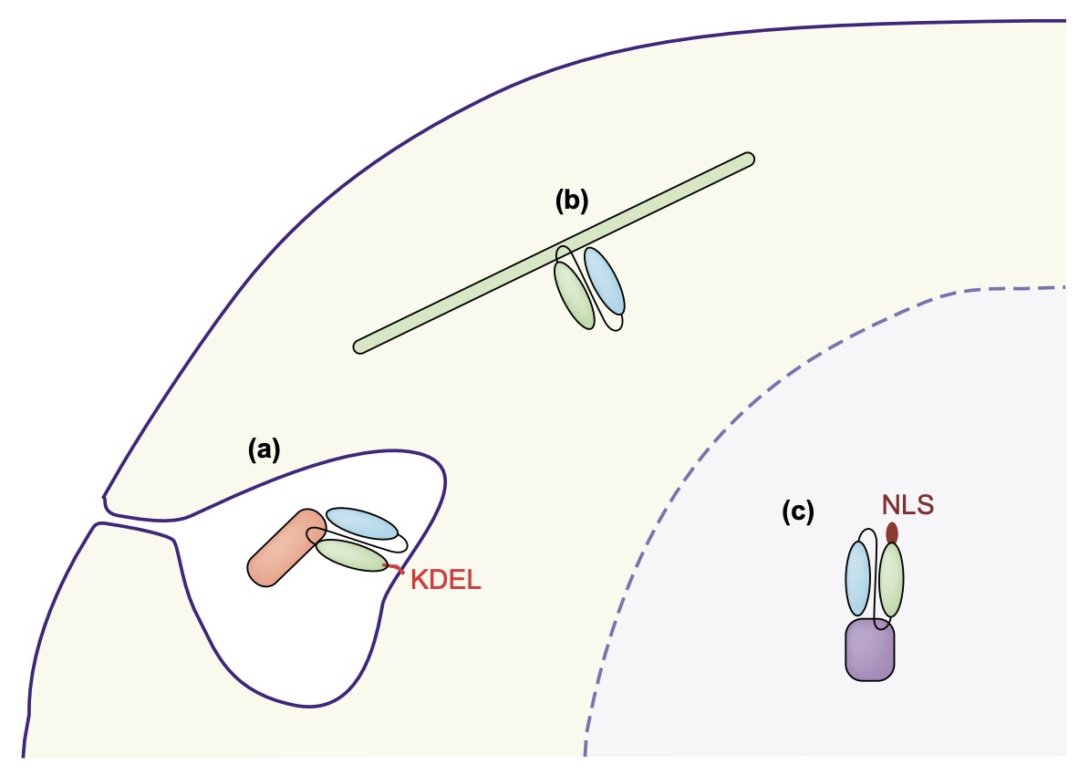
Targeting motifs such as NLS or mitochondrial targeting sequences are appended to direct intrabody localization. Such designs enable precise interrogation or modulation of compartment-specific protein networks. Certain intra-VHHs, when used as biosensors, have been localized to subcellular domains like clathrin-coated pits and actin-defined nanodomains. These findings highlight the need for careful engineering of localization sequences to ensure the intrabody reaches its target signaling hotspot, particularly when studying receptors with endosomal signaling dynamics.
Yeast and Bacterial Two-Hybrid Systems: Useful for initial selection under intracellular expression conditions
Mammalian Cell-Based Assays: Fluorescence imaging, co-localization, and functional readouts in relevant models
Imaging-Based Tools: FRAP, FRET, or chromobody analysis to validate binding dynamics, affinity, and specificity
Proteomic Profiling: Co-immunoprecipitation, IP-MS, or proximity-labeling approaches to assess target engagement and off-target interactions.
VHH intrabodies are genetically encoded single-domain binding reagents that can be used to visualize, stabilize, inhibit, relocalize, or degrade intracellular proteins in selected experimental systems. Their small size, monomeric architecture, and engineerability make them attractive for live-cell imaging and target perturbation, but intracellular performance is highly clone- and context-dependent. Key development challenges include cytosolic folding, expression level, subcellular localization, delivery, off-target binding, and functional validation in disease-relevant models.
Biointron’s VHH Antibody Discovery platform supports high-throughput selection of nanobodies tailored to research and clinical pipelines.
What is a VHH intrabody?
A VHH intrabody is a single-domain antibody derived from camelid heavy-chain antibodies, engineered to be expressed and function within the intracellular environment.
How are VHH intrabodies different from conventional antibodies?
Unlike full-length antibodies, VHHs are small, monomeric, and do not require disulfide bonds, allowing them to remain functional in the cytoplasm.
Can VHH intrabodies function without disulfide bonds?
Yes. Many VHHs retain activity without disulfide bonds, particularly when derived from llama sources or engineered for disulfide-free stability.
How are VHH intrabodies delivered into cells?
Delivery strategies include plasmid or mRNA transfection, viral vectors (e.g., lentivirus, AAV), and protein transduction using CPPs or physical methods.
What are the main challenges in using VHHs as intrabodies?
Challenges include ensuring cytoplasmic folding, optimizing delivery efficiency, and validating functional intracellular binding.
Are VHH intrabodies used in clinical research?
Most VHH intrabody applications remain preclinical, although VHH-based therapeutics more broadly have advanced into clinical use.
Wagner, T. R., & Rothbauer, U. (2020). Nanobodies Right in the Middle: Intrabodies as Toolbox to Visualize and Modulate Antigens in the Living Cell. Biomolecules, 10(12), 1701. https://doi.org/10.3390/biom10121701
Soetens, E., Ballegeer, M., & Saelens, X. (2020). An Inside Job: Applications of Intracellular Single Domain Antibodies. Biomolecules, 10(12), 1663. https://doi.org/10.3390/biom10121663
Messer, A., & Butler, D. C. (2020). Optimizing intracellular antibodies (intrabodies/nanobodies) to treat neurodegenerative disorders. Neurobiology of Disease, 134, 104619. https://doi.org/10.1016/j.nbd.2019.104619
Raynaud, P., Gauthier, C., Jugnarain, V., Jean-Alphonse, F., Reiter, E., Bruneau, G., & Crépieux, P. (2022). Intracellular VHHs to monitor and modulate GPCR signaling. Frontiers in Endocrinology, 13, 1048601. https://doi.org/10.3389/fendo.2022.1048601
Antibody discovery has become increasingly sequence-rich. Display technologies, ……
Biointron, a leading contract research organization specializing in antibody dis……
Post-translational modifications (PTMs) are chemical or structural changes made ……
Research recap on Antibody Engineering & Therapeutics Europe 2026. Antibody inno……