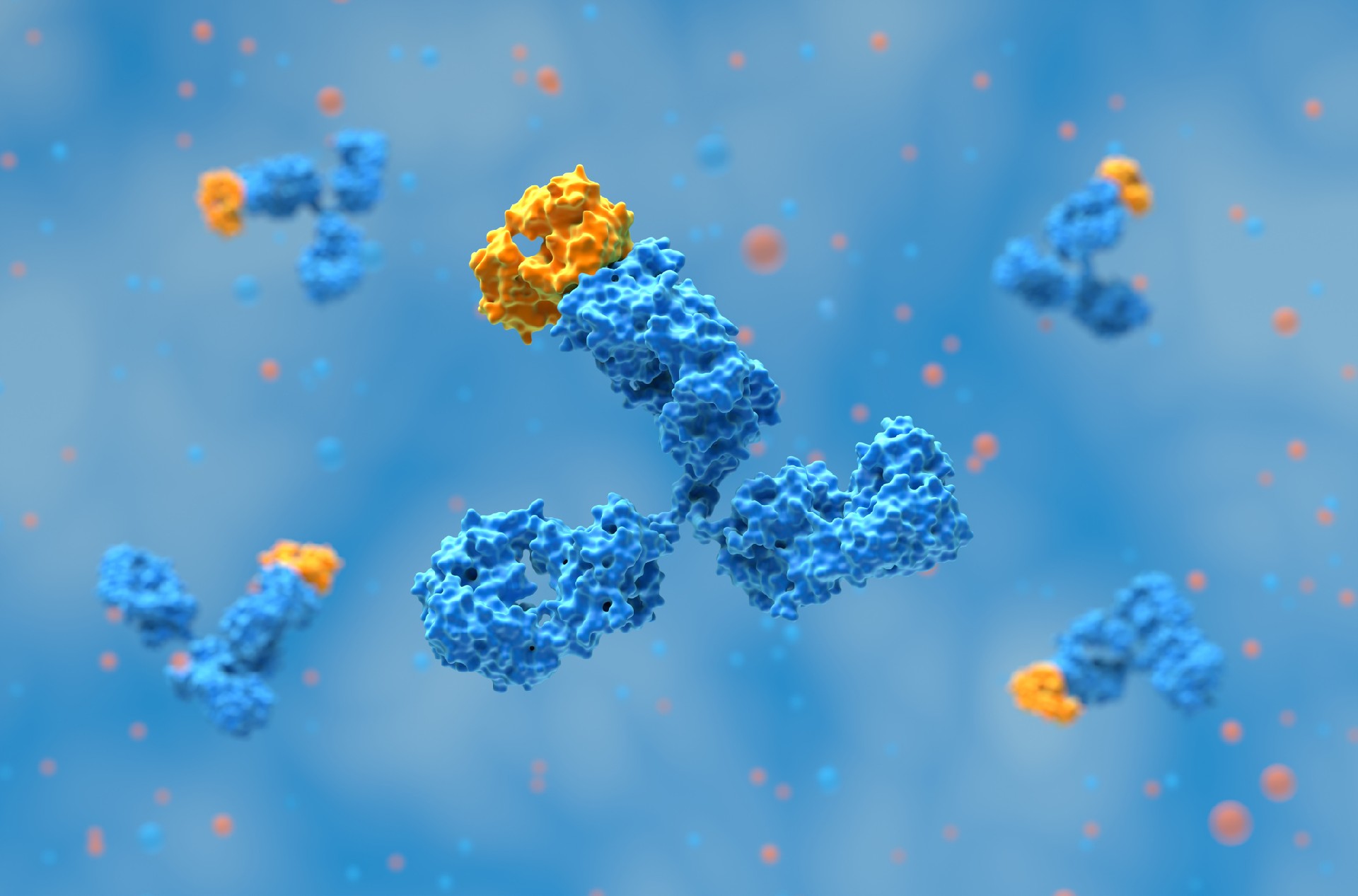
Hybridoma technology, developed by Köhler and Milstein in 1975, is a foundational method for producing monoclonal antibodies (mAbs). The approach involves fusing antibody-producing B lymphocytes with immortal myeloma cells to form hybridoma cells. These hybridomas combine the desirable traits of both parent cell types: the specificity and antigen recognition capability of B cells and the indefinite growth potential of myeloma cells. The resulting monoclonal antibodies are homogeneous, highly specific, and sensitive to a single epitope on a target antigen.
The monoclonal antibodies produced via hybridoma technology are applied extensively in diagnostics, imaging, therapeutics, and basic research. Their uniformity and specificity offer significant advantages over polyclonal antibodies, especially in clinical and industrial biotechnology contexts.
Hybridoma formation is achieved through the fusion of two distinct cell types:
B lymphocytes: Derived from the spleen of an immunized animal (typically a mouse), B cells are responsible for producing antigen-specific antibodies. However, they have a limited lifespan in vitro.
Myeloma cells: Immortal cancerous plasma cell lines, chosen for their ability to proliferate indefinitely. These cells are selected for lack of endogenous antibody secretion and for being HGPRT-deficient, which enables selective growth post-fusion.
The resulting hybridoma cell line inherits the immortality of the myeloma parent and the specific antibody-producing capability of the B lymphocyte. These cells can be cultured indefinitely in vitro, providing a continuous and consistent source of monoclonal antibodies.

The choice of fusion partners is critical for the success of hybridoma generation. B cells are harvested from immunized mice after sufficient antigen exposure and serum antibody generation. Myeloma cell lines are pre-treated with 8-azaguanine to induce HGPRT deficiency. This renders them sensitive to selection in hypoxanthine-aminopterin-thymidine (HAT) medium, as they cannot survive without functional salvage pathways for nucleotide synthesis.
Common myeloma cell lines used include SP2/0 and NS0. These are selected for being non-antibody-secreting and for their suitability in large-scale culture. The fusion efficiency and post-fusion viability of the hybridomas also depend on the quality and physiological state of the fusion partners.
The most frequently used method for fusing B lymphocytes with myeloma cells is polyethylene glycol (PEG)-mediated fusion. PEG promotes the fusion of plasma membranes, facilitating the formation of heterokaryons that can develop into viable hybridoma cells.

Alternatively, electrofusion is used, where cells are exposed to a controlled electric field to induce fusion. Electrofusion can provide higher efficiency and better control over fusion events.
Key parameters influencing fusion success include:
Ratio of B cells to myeloma cells
PEG concentration and exposure time
Cell viability and density during fusion
Following fusion, the mixed population includes unfused B cells, unfused myeloma cells, and hybridomas.
Post-fusion, the cell suspension is cultured in HAT medium for selective growth of hybridomas. HAT medium contains:
Hypoxanthine and thymidine: Support nucleotide synthesis via the salvage pathway.
Aminopterin: Inhibits the de novo pathway for nucleotide synthesis.
Only cells with a functional HGPRT gene, contributed by the B lymphocyte parent, can survive. Unfused myeloma cells, lacking HGPRT, die. Unfused B cells have a limited lifespan and do not proliferate. Consequently, only hybridomas survive and proliferate.
Clonal isolation of hybridomas is then carried out via limiting dilution, where single hybridoma cells are plated in individual wells. This ensures monoclonality of the resulting cultures and enables screening for specific antibody-producing clones.
Related: Hybridoma Sequencing
Clones are screened for production of the desired antibody using techniques like ELISA, which can detect antigen-specific antibodies in culture supernatants, thus allowing researchers to identify hybridomas producing antibodies with high affinity and specificity toward the target epitope.
Further analyses include:
Isotyping: Determines the antibody class (e.g., IgG, IgM).
Affinity testing: Assesses binding strength to the antigen.
Specificity evaluation: Confirms selectivity for the target without cross-reactivity.
Once the most suitable hybridoma clones are identified, they are expanded for large-scale antibody production.

Hybridoma sequencing is used to determine the nucleotide sequences of the variable regions in the heavy (VH) and light (VL) chains of monoclonal antibodies produced by hybridoma cells. This process provides a genetic blueprint for antibody structure, allowing for replication, modification, and optimization of monoclonal antibodies in research and therapeutic contexts.
A typical workflow involves these main steps:
RNA extraction from hybridoma cells.
Reverse transcription of RNA into complementary DNA (cDNA), often using RACE (Rapid Amplification of cDNA Ends) techniques to capture full-length variable regions.
PCR amplification of antibody gene segments.
Sequencing of the amplified gene products to obtain full VH and VL sequences.
This gene-level characterization offers several advantages over traditional amino acid sequencing, especially in cases where post-translational modifications or similar residues complicate mass spectrometric analysis. Nucleotide sequencing ensures accurate identification of each amino acid and maintains fidelity in antibody engineering efforts.
Sequencing enables the antibody to be preserved as a digital base sequence, eliminating dependence on hybridoma cell lines for continued production. This mitigates the risks of genetic drift, cell line loss, and variability associated with long-term hybridoma culture and cryopreservation.
Once the antibody gene sequence is known, recombinant expression systems can be used to produce the monoclonal antibody in large quantities with high consistency. This approach supports:
Expression optimization for improved yield and stability.
Batch-to-batch consistency critical for industrial and clinical applications.
Antibody engineering, including expression of fragments (e.g., Fab, scFv), isotype switching, chimerization, and humanization.
Additionally, hybridoma sequencing helps reduce experimental costs and improve the reproducibility of research by:
Facilitating the design of optimized purification and expression strategies.
Providing sequence data necessary for intellectual property filings.
Enabling affinity maturation through sequence-guided mutagenesis.
The final application of this data is in recombinant antibody production, which circumvents the animal immune system and reduces the risk of adverse immune responses in therapeutic settings. Recombinantly expressed antibodies offer superior control, scalability, and quality assurance for diagnostic and biopharmaceutical use.

Related: What is a Hybridoma?
Selected hybridoma clones are propagated using in vitro or in vivo methods:
In vitro culture in flasks or bioreactors offers a controlled and scalable environment, minimizing the risk of contamination and facilitating high-purity antibody recovery.
In vivo production involves injecting hybridoma cells into the peritoneal cavity of mice, where they produce ascitic fluid rich in antibodies. However, this method may introduce mouse-derived immunoglobulins and require additional purification.
Cryopreservation of hybridoma lines ensures long-term stability and reproducibility. Cells are frozen in cryoprotectant-containing media and stored in liquid nitrogen, enabling future use without re-immunization and fusion.
Related: Hybridoma Sequencing
Hybridoma-derived monoclonal antibodies have significant utility in the biotechnology and CRO sectors:
Diagnostics: Detection of pathogens, toxins, and biomarkers (e.g., hCG in pregnancy tests).
Therapeutics: Use in targeted cancer therapies (e.g., rituximab), autoimmune disorders, and infectious diseases.
Radioimmunodetection and radioimmunotherapy: mAbs labeled with isotopes for imaging and treatment of tumors.
Serological applications: Blood group typing, pathogen strain differentiation.
Contract research organizations (CROs) leverage hybridoma technology for custom antibody development, validation, and supply chain continuity in drug discovery pipelines.
Despite its widespread use, hybridoma technology has limitations:
Species restriction: Primarily based on murine systems, raising concerns about immunogenicity in humans.
Contamination risk: Cell cultures are susceptible to viral or microbial contamination.
Fusion efficiency: Low overall yield; <2% of cells form viable hybridomas.
Viral risk: Potential transmission of murine viruses to humans, even after purification.
Time and cost: Hybridoma generation is labor-intensive and takes 6–9 months.
To overcome these limitations, newer techniques have emerged:
Chimeric and humanized antibodies: Reduce immunogenicity through genetic engineering.
Transgenic mice: Engineered to express human immunoglobulin genes for human mAb production.
Recombinant DNA methods: Use of phage display for high-throughput antibody generation.
Bispecific antibodies: Target two different antigens simultaneously, enhancing therapeutic value.
These alternatives enable the development of next-generation antibody therapeutics with improved pharmacokinetics, safety, and efficacy.
References:
Mitra, S., & Tomar, P. C. (2021). Hybridoma technology; advancements, clinical significance, and future aspects. Journal of Genetic Engineering & Biotechnology, 19, 159. https://doi.org/10.1186/s43141-021-00264-6
Antibody discovery has become increasingly sequence-rich. Display technologies, ……
Biointron, a leading contract research organization specializing in antibody dis……
Post-translational modifications (PTMs) are chemical or structural changes made ……
Research recap on Antibody Engineering & Therapeutics Europe 2026. Antibody inno……