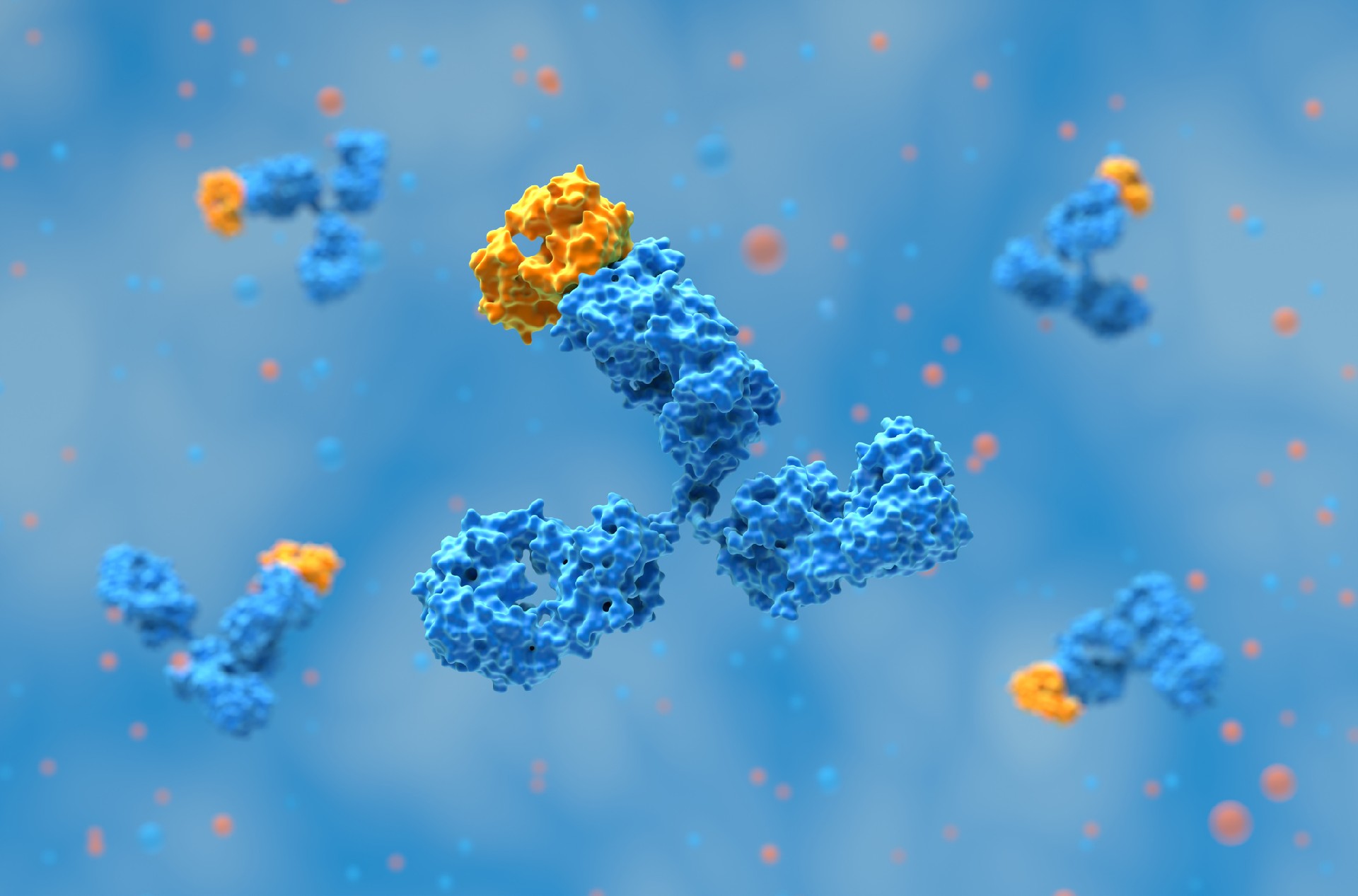
VHH antibodies, also called single-domain antibodies or nanobodies, are the smallest functional fragments of antibodies capable of binding antigens. They originate from the heavy-chain-only antibodies found in camelids such as llamas and camels. Unlike conventional antibodies, which require both heavy and light chains for antigen recognition, VHHs operate as a single domain.
This structural simplicity gives them distinct advantages in terms of their size, stability, and solubility.
A typical VHH domain is composed of about 120–135 amino acids. Despite its smaller size compared to an IgG molecule, it maintains the classical immunoglobulin fold. The structure can be divided into:
Four framework regions (FR1–FR4) that stabilize the domain.
Three complementarity-determining regions (CDR1–CDR3) that form the antigen-binding site.
The most notable adaptation is the CDR3 loop, which is longer and more flexible than in conventional VH domains. This compensates for the absence of a paired light chain by allowing VHHs to access recessed or cryptic epitopes that standard antibodies cannot easily reach.
Several physicochemical properties distinguish VHHs from full-length antibodies:
Size: At roughly 15 kDa, a VHH is one-tenth the size of a full IgG (~150 kDa). This structure enables deeper tissue penetration, improved biodistribution, and access to areas such as the central nervous system.
Stability: VHHs can resist extreme conditions, including high temperatures, acidic or alkaline pH, and denaturing environments. This resilience makes them well-suited for therapeutic delivery in variable physiological conditions as well as for field diagnostics where cold-chain storage is limited.
Solubility: Framework substitutions enhance hydrophilicity, minimizing aggregation that can compromise antibody performance. High solubility also supports their use in high-concentration formulations, which are required for certain therapeutic routes like inhalation or topical delivery.
Aggregation resistance: VHHs resist self-association, which improves long-term stability and reduces the risk of immunogenic responses caused by aggregates. This property is particularly important in biopharmaceutical development, where manufacturability and reproducibility are critical.
Shelf-life: Due to their intrinsic stability, VHHs can be stored for extended periods without loss of activity. This makes them ideal for use in diagnostic kits, biosensors, and other applications requiring reliability over time.
These features make VHHs attractive for applications in imaging, biosensors, and intracellular targeting, where structural robustness is critical.
The VHH domain is one of the smallest naturally occurring antigen-binding units in vertebrates, comparable in size to the IgNAR variable domain (vNAR) from sharks.
Despite arising independently, both domains exhibit single-domain solubility, elongated CDR3 loops, and high thermal stability, which is an example of convergent evolution toward compact yet functional antigen recognition systems.
The framework regions of VHHs contain adaptations that ensure solubility and folding stability without the support of a light chain. In particular, the FR2 region shows hydrophilic substitutions at key positions such as 44, 45, and 47.
In conventional VH domains, these positions are hydrophobic to stabilize pairing with VL domains. In VHHs, however, hydrophilic residues prevent unwanted aggregation and allow the domain to fold independently. This explains how heavy-chain-only antibodies can remain fully functional in camelids.
A defining feature of VHH domains is the set of hallmark amino-acid substitutions within framework region 2 (FR2), notably V37F/Y, G44E, L45R, and W47G (Kabat numbering).
These residues occupy positions that, in conventional VH domains, mediate hydrophobic interactions with the light-chain variable domain (VL).
In the absence of a VL partner, camelid VHHs replace these hydrophobic contacts with polar side chains, greatly improving solubility and folding stability as independent single domains.
This structural adaptation allows VHHs to remain monomeric and functional without forming aggregates, a prerequisite for their biotechnological robustness.
The antigen-binding site of a VHH is shaped primarily by the CDR loops, with the CDR3 loop playing a dominant role. Its length and flexibility give VHHs the ability to:
Extended CDR3 loop: The long and variable CDR3 often folds into shapes that can penetrate enzyme clefts, receptor pockets, or other hidden epitopes. This makes VHHs capable of binding targets such as G protein–coupled receptors (GPCRs), viral surface proteins, or enzyme active sites that are inaccessible to full-length IgGs.
Binding orientation: Unlike IgGs, which usually interact with broader, exposed surfaces on antigens, VHHs can adopt orientations that reach recessed or conformationally complex regions. This orientation flexibility helps explain their success in targeting protein–protein interaction sites, a long-standing challenge in drug discovery.
High affinity despite small size: Even though they are a fraction of the size of conventional antibodies, VHHs achieve nanomolar to picomolar binding affinities. This is often due to the way the CDR3 loop pairs with the supporting CDR1 and CDR2 regions to form a highly complementary binding surface.
Structural diversity: The diversity of CDR3 length and conformation across camelid repertoires increases the likelihood of discovering VHHs against “difficult” antigens, including toxins, viral epitopes, and membrane proteins.
Unlike full IgGs that often engage flat or exposed epitopes, VHHs specialize in binding unique structural regions. This property makes them valuable for targeting GPCRs, enzymes, and protein–protein interaction sites.
High-resolution crystal structures have shown that VHH paratopes exhibit remarkable conformational versatility.
Depending on the CDR loop arrangement, the binding surface may be penetrative, flat, convex, or concave, enabling interactions with a broad range of antigenic topologies, from enzyme active sites to protein–protein interfaces.
This diversity stems mainly from the elongated CDR3 loop, which can fold back onto the framework, form intradomain disulfides with CDR1, or extend outward to access deep cavities.
Such plasticity distinguishes VHHs from classical antibodies, whose paratopes are generally limited to flatter surfaces.
Crystallographic studies of VHH-antigen complexes have confirmed how framework residues and extended CDR3 loops determine binding modes. These structures highlight the role of VHHs in accessing targets that larger antibodies cannot reach.
In addition, computational tools now support nanobody structure prediction and design. Programs such as AlphaFold, NanoBodyBuilder, and Rosetta enable researchers to model VHH domains, refine stability, and optimize affinity. These approaches accelerate the design of improved VHHs before moving into experimental validation.
The unique VHH antibody structure directly supports antibody engineering. Common design strategies include:
Multivalent constructs: tandem VHHs or bispecific designs that increase functional potency.
Fc fusions or albumin-binding fusions: added domains that extend half-life and improve pharmacokinetics.
Intrabodies: VHHs expressed inside cells to modulate intracellular protein function.
These engineering approaches demonstrate how structural insights translate into practical formats for drug development and research tools.
VHH domains are characterized by exceptional thermostability, with melting temperatures commonly between 50 °C and 80 °C.
Many VHHs regain functional conformation after heating and cooling cycles, which underpins their use in diagnostics and biosensors that must tolerate temperature fluctuations.
However, full reversibility is not universal: thermal unfolding and aggregation tendencies can vary among clones, particularly at high protein concentrations.
For therapeutic candidates, temperature-dependent aggregation assays are often employed early in development to identify variants with optimal stability and manufacturability profiles.
The structure of VHH antibodies explains why they are considered one of the most versatile antibody formats in modern science. Their single-domain design, long CDR3 loop, and framework adaptations combine to produce stability, solubility, and high-affinity binding.
These features enable applications ranging from therapeutic antibodies and nanobody-based drugs to diagnostic platforms and biosensors. By linking molecular design to functional performance, the study of VHH antibody structure continues to guide innovation in antibody engineering and biomedical research.
References:
Hoey, R. J., Eom, H., & Horn, J. R. (2019). Structure and development of single domain antibodies as modules for therapeutics and diagnostics. Experimental Biology and Medicine, 244(17), 1568. https://doi.org/10.1177/1535370219881129
Two popular topics discussed at the 2026 BIO International Convention were devel……
AI is changing antibody discovery, but model performance depends on the quality ……
Antibody discovery has become increasingly sequence-rich. Display technologies, ……
Biointron, a leading contract research organization specializing in antibody dis……